Copyright
©2009 Baishideng.
World J Gastrointest Surg. Nov 30, 2009; 1(1): 62-64
Published online Nov 30, 2009. doi: 10.4240/wjgs.v1.i1.62
Published online Nov 30, 2009. doi: 10.4240/wjgs.v1.i1.62
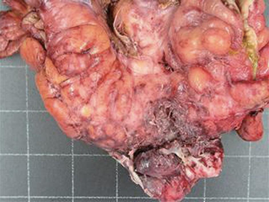
Figure 1 Surgical specimen: Sigmoid colon mass (80 mm × 30 mm) with abscess formation.
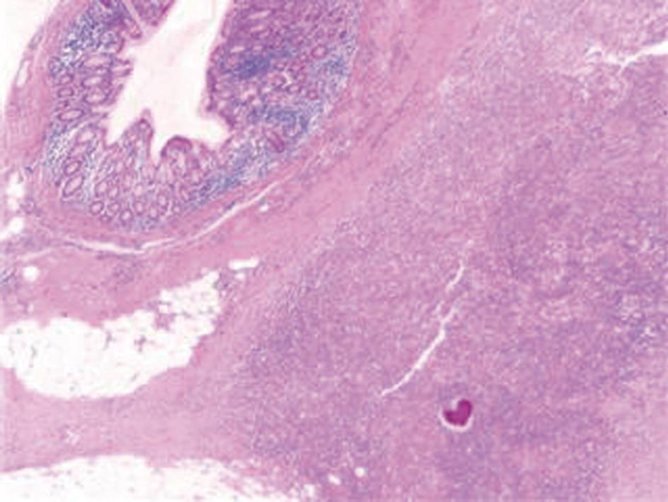
Figure 2 Histology: A colony of Actinomyces is seen within the pericolonic inflammatory tissue.
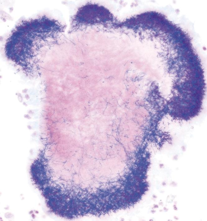
Figure 3 Gram-positive staining of an Actinomyces colony.
- Citation: Privitera A, Milkhu CS, Datta V, Rodriguez-Justo M, Windsor A, Cohen CR. Actinomycosis of the sigmoid colon: A case report. World J Gastrointest Surg 2009; 1(1): 62-64
- URL: https://www.wjgnet.com/1948-9366/full/v1/i1/62.htm
- DOI: https://dx.doi.org/10.4240/wjgs.v1.i1.62


