Copyright
©2009 Baishideng.
World J Gastrointest Surg. Nov 30, 2009; 1(1): 59-61
Published online Nov 30, 2009. doi: 10.4240/wjgs.v1.i1.59
Published online Nov 30, 2009. doi: 10.4240/wjgs.v1.i1.59
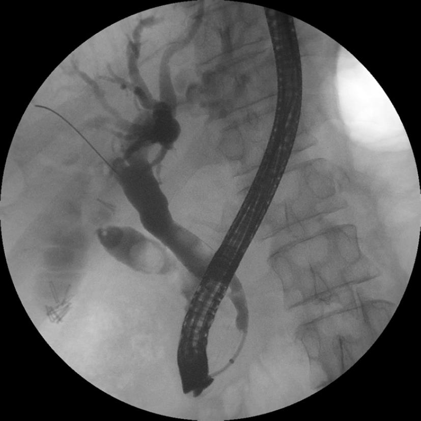
Figure 1 Fluoroscopic image obtained during ERCP showing filling defects (stones) in the common hepatic duct and cystic duct remnant.
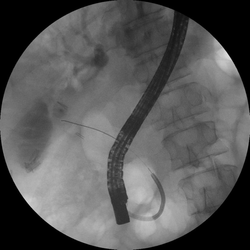
Figure 2 A guidewire is placed in the cystic duct remnant under direct cholangioscopic guidance.
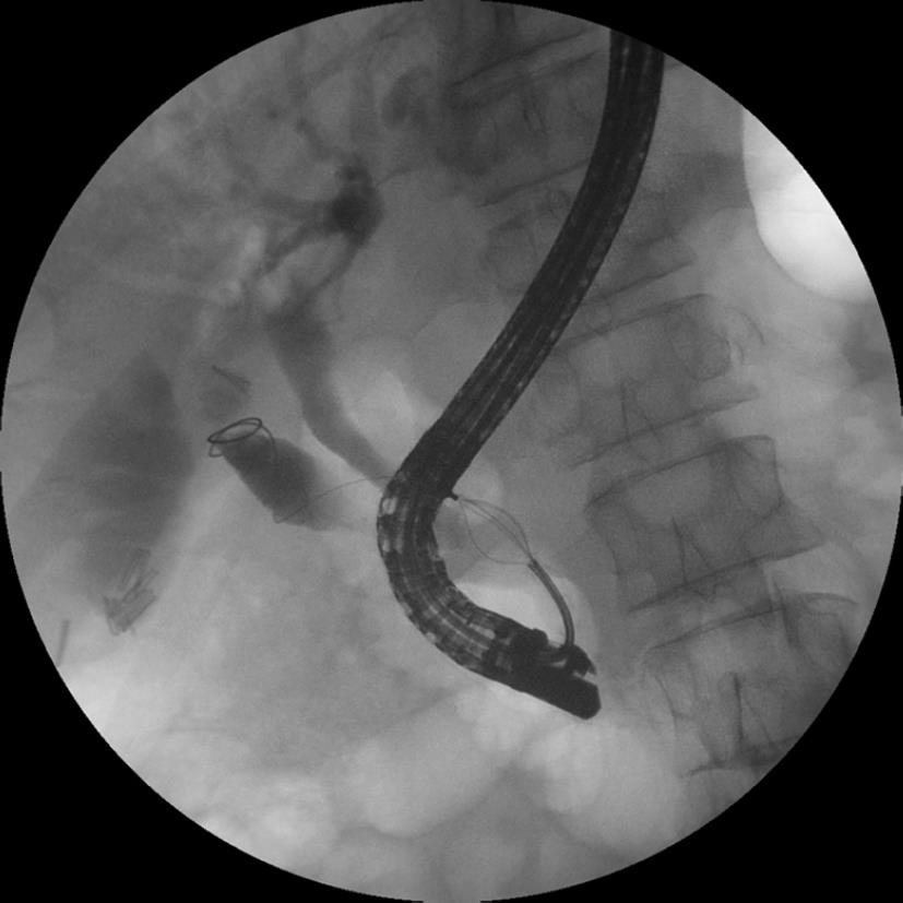
Figure 3 A guidewire is coiled in the cystic duct remnant.
A basket passed over the guidewire is holding a large radiolucent (cholesterol) stone for removal.
- Citation: Parsi MA. Peroral cholangioscopy-assisted guidewire placement for removal of impacted stones in the cystic duct remnant. World J Gastrointest Surg 2009; 1(1): 59-61
- URL: https://www.wjgnet.com/1948-9366/full/v1/i1/59.htm
- DOI: https://dx.doi.org/10.4240/wjgs.v1.i1.59


