Minireviews Open Access
Copyright ©The Author(s) 2023. Published by Baishideng Publishing Group Inc. All rights reserved.
World J Diabetes. Jun 15, 2023; 14(6): 795-807
Published online Jun 15, 2023. doi: 10.4239/wjd.v14.i6.795
Diabetes and cognitive decline: Challenges and future direction
Norhamidar Ab-Hamid, Idris Long, Biomedicine program, School of Health Sciences, Health Campus, Universiti Sains Malaysia, Kubang Kerian, Kota Bharu 16150, Kelantan, Malaysia
Norsuhana Omar, Che Aishah Nazariah Ismail, Department of Physiology, School of Medical Sciences, Health Campus, Universiti Sains Malaysia, Kubang Kerian, Kota Bharu 16150, Kelantan, Malaysia
ORCID number: Norsuhana Omar (0000-0002-1975-6487); Che Aishah Nazariah Ismail (0000-0003-1723-3575); Idris Long (0000-0002-6828-1908).
Author contributions: Long I and Ab-Hamid N prepared the manuscript; Ismail CAN and Omar N proofread this manuscript.
Supported by the Fundamental Research Grant Scheme (FRGS) Ministry of Higher Education Malaysia, No. FRGS/1/2020/SKK0/USM/03/5.
Conflict-of-interest statement: All authors report having no relevant conflicts of interest for this article.
Open-Access: This article is an open-access article that was selected by an in-house editor and fully peer-reviewed by external reviewers. It is distributed in accordance with the Creative Commons Attribution Noncommercial (CC BY-NC 4.0) license, which permits others to distribute, remix, adapt, build upon this work non-commercially, and license their derivative works on different terms, provided the original work is properly cited and the use is non-commercial. See: https://creativecommons.org/Licenses/by-nc/4.0/
Corresponding author: Idris Long, PhD, Academic Research, Senior Lecturer, Biomedicine program, School of Health Sciences, Health Campus, Universiti Sains Malaysia, Jalan Hospital USM, Kubang Kerian, Kota Bharu 16150, Kelantan, Malaysia. idriskk@usm.my
Received: December 25, 2022
Peer-review started: December 25, 2022
First decision: January 3, 2023
Revised: February 7, 2023
Accepted: May 6, 2023
Article in press: May 6, 2023
Published online: June 15, 2023
Processing time: 171 Days and 17.8 Hours

Abstract

There is growing evidence that diabetes can induce cognitive decline and dementia. It is a slow, progressive cognitive decline that can occur in any age group, but is seen more frequently in older individuals. Symptoms related to cognitive decline are worsened by chronic metabolic syndrome. Animal models are frequently utilized to elucidate the mechanisms of cognitive decline in diabetes and to assess potential drugs for therapy and prevention. This review addresses the common factors and pathophysiology involved in diabetes-related cognitive decline and outlines the various animal models used to study this condition.

Key Words: Diabetes mellitus; Insulin signaling; Macrovascular disease; Microvascular disease; Animal models; Cognitive decline; Pathophysiology

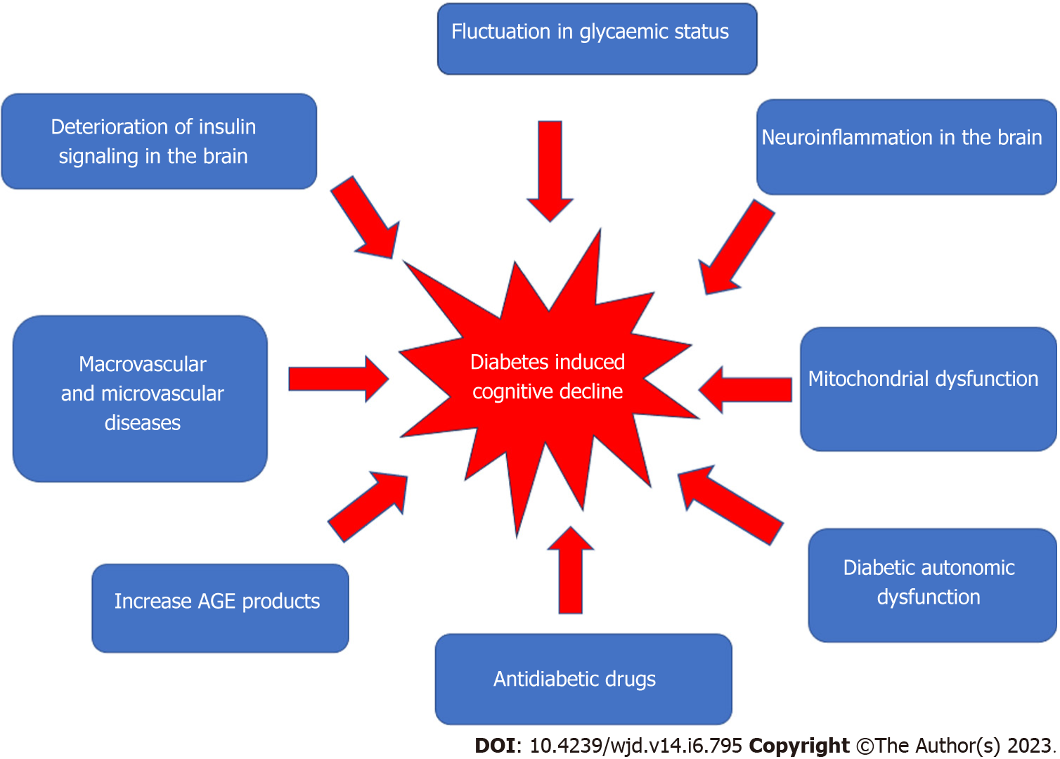
Core Tip: Diabetes can induce cognitive decline, a phenomenon attributed to fluctuations in glycemic status, macrovascular and microvascular disease, deterioration of insulin signaling, neuroinflammation, mitochondrial dysfunction, increases in advanced glycation end products, the effects of drugs used to treat diabetes, and diabetic autonomic dysfunction. Various animal models have been constructed to examine the pathophysiology of diabetes-induced cognitive decline.


INTRODUCTION

According to current data from the International Diabetes Federation (IDF), more than 1 in 10 ten persons has diabetes. Diabetes prevalence among adults (20 to 79 year of age) has more than quadrupled from 151 million (4.6% of the global population) in 2000 to 537 million (10.5% of the global population) in 2021. More shockingly, it is estimated that without changes to intervention strategy, 643 million people (11.3% of the world population) will develop diabetes by 2030[1].

Diabetes complications are primarily attributed to vascular and metabolic factors associated with the disease. Among the major complications are cardiovascular disease, stroke, peripheral artery disease, nephropathy, retinopathy, neuropathy, dental disease, and immunocompromise. These are also expected to become increasingly pervasive, affecting both the local and global burden of illness[2]. Diabetes is a systemic disease, as it can affect nearly every body system. For instance, diabetes can disrupt proper cardiovascular, gastrointestinal, immune, or nervous function. The functional impairment of the peripheral nervous system can lead to diabetic foot and, in the worst cases, amputation and associated physical disability. Diabetic retinopathy can lead to loss of vision and blindness. A wide range of cognitive dysfunction can also occur as a consequence of diabetes, and can manifest as mild cognitive impairment (MCI) to dementia[3].

There is an increased risk of cognitive decline and dementia in patients with diabetes[4]. This has major implications for patient care, particularly in older adults with dementia or pre-dementia with cognitive impairment, which are the most typical manifestations in communities worldwide[4]. The stages of diabetes-associated cognitive decline depend on the type of diabetes and also the patient’s age. For type 1 diabetes (T1DM), the impairment of cognitive function progresses with age. However, for type 2 diabetes (T2DM), there are three stages of cognitive function loss, including diabetes-associated cognitive decline, MCI, and in the final stage, dementia[2].

Several investigations have revealed that patients with T1DM may suffer from severe impairments in information processing, psychomotor efficiency, attention, visuoconstruction, and mental flexibility[5]. However, T2DM has been associated more with problems in executive function, psychomotor speed, and memory. As a result, older diabetic patients often have slower walking pace, poorer coordination, a higher chance of falling, and more fractures, all of which can affect quality of life. In addition, executive dysfunction has been linked to the incapacity to carry out daily tasks.

The effects of diabetes on brain function and cognitive decline have received little attention in academia. However, a study using brain magnetic resonance spectroscopy discovered various metabolic criteria for dementia in diabetic patients and established new links between dementia and diabetes. This study also found extremely low levels of N-acetyl aspartate (which affects neuronal integrity), high levels of myoinositol, high levels of excitatory neurotransmitters (e.g., glutamate and glycine), and low levels of inhibitory neurotransmitters [e.g., gamma-aminobutyric acid (GABA)], which has been linked to pain perception problems in diabetic patients[6]. Diabetes also causes brain atrophy, myelin degradation, and vacuole dispersion throughout the white matter of the brain in rats[7]. Diabetic patients are also thought to have irregularities in the metabolism of neurotransmitters in the brain, which leads to neuronal dysfunction and destruction, eventually contributing to the development of dementia.

Research regarding the actions of insulin has mainly focused on peripheral diseases rather than brain function[8]. However, insulin has been shown to play a role in cognition and neuroprotection in brain. Insulin also has an indirect effect on brain function by acting on peripheral tissue. Many circulating mediators that fluctuate due to obesity and diabetes can pass across the blood-brain barrier (BBB) and contribute to dysfunction in neurons, astrocytes, and microglia[7]. Nonetheless, the mechanism of diabetes-induced cognitive decline is still uncertain. Interestingly, this condition shares many cellular and molecular pathways with Alzheimer’s disease (AD), the most common form of dementia[9]. Here, we describe the putative pathophysiology of diabetes that may contribute to cognitive decline and review diabetic animal models used to study this condition. Finally, we discuss the obstacles and future directions for elucidating the diabetes-related mechanisms associated with cognitive decline.

FACTORS AND PATHOPHYSIOLOGY OF DIABETES INDUCES A COGNITIVE DECLINE

Figure 1 shows the factors that may contribute to the development of diabetes-induced cognitive decline.

Figure 1
Figure 1 Multifactorial pathophysiology of diabetes-induced cognitive decline.
Macrovascular and microvascular diseases

Many vascular, metabolic, and psychosocial factors have been linked to diabetic-induced cognitive decline. Vascular disease, including hypertension and dyslipidemia, has been linked to an increased risk of stroke; in diabetic patients, this risk is estimated to be 115% higher for every 1% increase in glycated hemoglobin (HbA1c)[10]. Furthermore, cardio- or cerebrovascular abnormalities in the brain can lead to cognitive decline and dementia. Patients with T1DM who frequently have cognitive difficulties may have both subclinical and overt cerebrovascular disease[3]. T2DM patients with elevated plasma triglycerides and higher cholesterol levels have been demonstrated to have poorer cognitive function[11]. Studies have also revealed a relationship between cognitive dysfunction and hypertension in T2DM[6,7]. However, due to inconsistent findings from observational studies in the general population[8,9], the roles of dyslipidemia and hypertension in the development of cognitive decline in diabetics is still uncertain and needs further investigation.

Microvascular dysfunction also has been associated with cognitive decline[3,12]. Chronic hyperglycemia increases the risk of microvascular dysfunction, which can affect many organs, including the eye (retinopathy), kidney (nephropathy), and nerves (neuropathy). There is also a positive correlation between the development of cognitive decline and the presence of nephropathy and/or retinopathy[13]. Retinopathy has been linked to cognitive decline in adult diabetic patients as it is thought to affect intelligence, attention/concentration, and information processing[14].

Hyperglycemia

Hyperglycemia has been linked to cognitive decline[15], and can affect cognitive function in the long and short term time. Hyperglycemia has been shown to correlate with impaired working memory, attention, and depression. Acute variations in blood glucose have a negative effect on cognitive performance, and glycemic control improvement is advantageous for regulating cognitive function. It does not cause microvascular structural changes, but has been linked to regional cerebral blood flow or osmotic shifts across the neuronal membrane[16]. In contrast, chronic hyperglycemia affects cognitive function through the production of advanced glycosylation end-products (AGEs), formation of senile plaques and neurofibrillary tangles, and cerebral microvascular disease[17]. A reduction in white matter volume has also been connected to diminished executive function and a reduction in the processing of information[18].

The negative impact of hyperglycemia on cognitive impairment was validated in a zebrafish study in which T1DM was induced with injection of streptozotocin (STZ)[19]. It was discovered that exposing zebrafish to water-diluted glucose for 14 d caused sustained memory impairment accompanied by an increase in acetylcholinesterase activity. On the other hand, galantamine therapy reversed the memory-damaging effects of hyperglycemia. These findings revealed a link between acetylcholinesterase activity and cognitive impairment in T1DM patients[19].

However, cross-sectional studies investigating the association of chronic hyperglycemia (as evidenced by HbA1c) and cognitive decline in people with T2DM have yielded inconclusive results[20,21]. However, this association is apparent in older patients, as the improvement in glycemic control also improves cognitive function[22]. One study demonstrated that treatment of T2DM with either rosiglitazone or glibenclamide (glyburide) improved working memory over 24 wk[16]. Metformin has also been shown to reduce the risk of cognitive impairment in diabetic patients[23], but other evidence suggests that it may increase the risk[24] or have no effect[25]. Treatment with metformin may reduce tau phosphorylation as well as interleukin-1ß-mediated activation of the phosphokinases Akt and mitogen-activated protein kinase (MAPK). Furthermore, it can inhibit the mitochondrial respiratory chain, increasing cyclic adenosine monophosphate (AMP) and activating protein kinase A and AMP-activated protein kinase (AMPK)[26]. AMPK activation has been shown to improve memory and learning in female animal models[27]. However, when the evidence from observational studies and randomized controlled trials is combined, it seems that hyperglycemia and glucose excursions are both weakly associated with poorer cognitive function in T2DM patients[28]. As a result, further research is needed regarding hyperglycemia as a potentially modifiable risk factor for cognitive decline in diabetes.

Hypoglycemia

The presence of hypoglycemic episodes in diabetic patients has also been linked to cognitive decline and an increased risk of dementia[17,18]. The human brain, which accounts for 20% of the body’s metabolic consumption, has a greater need for glucose as a fuel source than other parts of the body. As a result, if the brain is temporarily depleted of glucose, cognitive and emotional functions are impaired. If left untreated, neuroglycopenia can lead to coma, seizure, or brain damage. There is evidence that repeated severe hypoglycemia in patients with early-onset diabetes can contribute to slower mental development and lower intellectual quotient (IQ)[29]. While the cerebral effects of severe hypoglycemia in adults are still not fully clear, insulin-dependent diabetic adults with repeated episodes of severe hypoglycemia performed worse on neuropsychological tests than diabetic patients who had never experienced severe hypoglycemia[30,31]. Another study found a weak link between the reported frequency of severe hypoglycemia and IQ decrement, lower levels of current IQ, and slowed variable reaction times[32]. However, this study found no cognitive differences between diabetic patients receiving intensive insulin therapy with severe hypoglycemia and those receiving conventional therapy[33]. The impact of several episodes of severe hypoglycemia between the ages of 5 year and 15 year is considered mild among young adults dependent on insulin. Strict glycemic control is thought to have a significant benefit in reducing target organ damage and slowing the progression of nephropathy, retinopathy, and neuropathy; however, it increases the risk of severe hypoglycemia. Further research on hypoglycemia and cognitive decline is needed to assist diabetic patients and their physicians in making the best treatment decisions.

Hyperinsulinemia

Hyperinsulinemia caused by endogenous insulin hypersecretion is common in the early stages of T2DM as a result of insulin resistance (IR). Hyperinsulinemia in adults without diabetes is associated with poorer cognitive function[26-28] and an increased risk of AD[34]. When compared to normal patients, patients with moderate to severe AD had higher levels of insulin in plasma but lower levels in cerebrospinal fluid[35]. Insulin therapy, both intravenous and intranasal, has been shown to improve cognitive function in AD patients[36]. Insulin injection into the cerebral ventricles of rats has also been shown to improve memory in a study that demonstrated an insulin signaling defect similar to that found in peripheral tissues could also occur in the hippocampus, resulting in functional insulin deficiency and cognitive decline[37]. Rosiglitazone can prevent disruption in memory tasks and reduce β-amyloid protein in the brain in transgenic mice that overexpress human amyloid precursor protein and develop AD pathology[38,39]. However, more research is needed to determine the link between hyperinsulinemia and cognitive decline.

Peripheral and cerebral insulin resistance

Insulin is released into the circulation by the pancreas and can pass the BBB via a carrier-facilitated process. The BBB comprises ependymal and endothelial cells, and the blood-cerebral spinal fluid barrier has insulin-binding sites that allow insulin to pass through[7]. Insulin receptors are found in the hypothalamus, prefrontal cortex, and hippocampus, among other central nervous system (CNS) sites[9]. The activation of hippocampal insulin receptors is thought to mediate insulin-induced cognitive improvement in healthy mammalian brains by facilitating long-term hippocampal potential (LTP), which is linked to learning and memory, as well as by increasing the expression of N-methyl-D-aspartate (NMDA) receptor[10] (Figure 2). Insulin also regulates the production of other neurotransmitters involved in learning and memory, including acetylcholine, norepinephrine, and adrenaline[3], and stimulates the accumulation of GABA-A receptors on the postsynaptic membrane[11]. A transient surge in peripheral insulin is thought to cause an increase in CNS insulin, which reaches the brain.

Figure 2
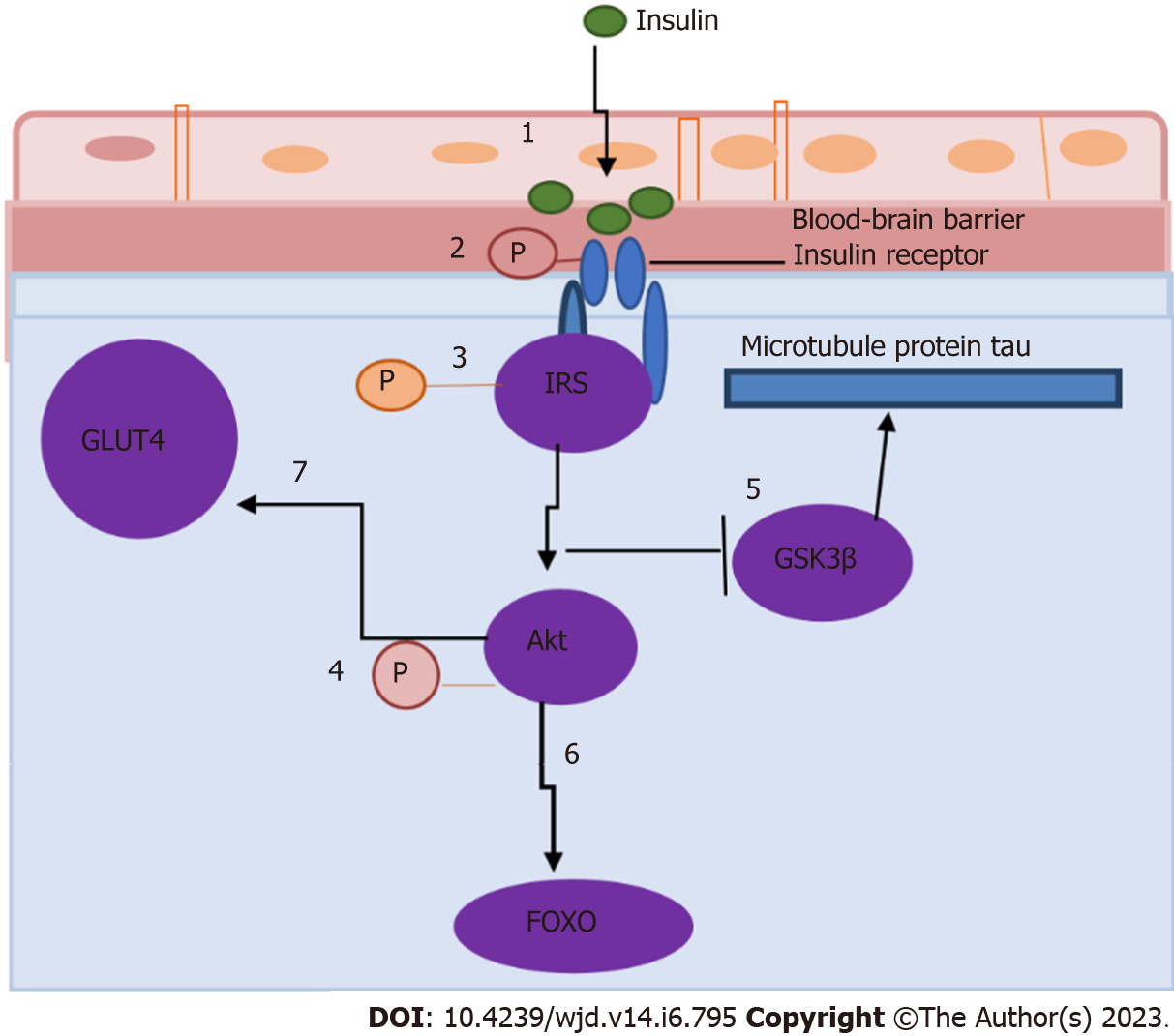
Figure 2 Insulin receptor signaling in the hippocampus (adapted from Biessels and Reagan[40], 2015). Cerebral insulin resistance causes downregulation of insulin transporters at the blood-brain barrier, limiting the amount of insulin that can enter the brain (step 1), decreasing the expression and/or activity of insulin receptors (step 2), modulating the phosphorylation state of insulin receptor substrates (step 3), phosphorylation of Akt (step 4), affecting several downstream components in the insulin signaling cascade (including glycogen synthase kinase 3β) (step 5), regulating the phosphorylation state of the microtubule protein tau and forkhead box O family of transcription factors (step 6), and impairing the trafficking of GLUT4 to the plasma membrane of the brain (step 7). GSK3β: Glycogen synthase kinase 3β; FOXO: Forkhead box O.

In healthy individuals, insulin binds to insulin receptor α-subunits and stimulates the tyrosine kinase domain of βsubunits, resulting in autophosphorylation. This autophosphorylation has the potential to activate the phosphoinositide 3-kinase (PI3K)-Akt (also known as PKB) signaling pathway. Akt molecules (Akt1, Akt2, and Akt3) are serine/threonine kinases that are activated by PI3K in response to growth factors and other cellular stimuli. In the brain, Akt mediates the translocation of glucose transporter type 4 (GLUT 4; also known as SLC2A4) to the plasma membrane. Akt also phosphorylates and inactivates the forkhead box O (FOXO) transcription factor family and glycogen synthase kinase 3β (GSK3β), reducing GSK3β’s ability to phosphorylate the microtubule-associated protein tau.

Chronic peripheral hyperinsulinemia induced by diabetes, obesity, or hyperlipidemia produces peripheral IR associated with the brain’s functional and structural changes. It also contributes to the dysregulation of insulin signaling in the brain and the development of cerebral IR. Cerebral IR causes the downregulation of insulin transporters at the BBB, limiting the amount of insulin that can enter the brain, decreasing the expression and/or activity of insulin receptors, and modulating the phosphorylation state of insulin receptor substrates such as Akt[7]. T2DM patients have lower Akt activation in their adipocytes and skeletal muscle, leading to many damaging effects on neuronal and glial cells[40]. Lower Akt activation affects several downstream components in the insulin signaling cascade, including GSK3β, which regulates the phosphorylation state of the microtubule protein tau and FOXO family of transcription factors. As a result, trafficking of GLUT4 to the plasma membrane is impaired. In addition, memory problems, diminished neuroprotective effects, and impaired synaptic transmission may result from cerebral IR, all of which may also contribute to the development of neurodegenerative illness[20]. However, the mechanisms underlying the relationships between systemic metabolic and vascular consequences of peripheral IR and cerebral IR are still largely undefined. Observational studies in humans are unlikely to fully elucidate the complex interplay between local and systemic factors of IR in the brain and periphery with respect to the mechanism of diabetes-induced cognitive decline[41,42].

Mitochondrial dysfunction

Mitochondria are involved in oxidative respiration, energy metabolism, free radical production, and apoptosis among other physiological processes[43]. The brain has a high energy requirement, and as such it is particularly sensitive to mitochondrial dysfunction. Mitochondria play an important role in anti-aging and neurodegenerative disease prevention[44]. The pathogenesis of diabetes and many neurodegenerative diseases includes mitochondrial dysfunction, attributed to the production of reactive oxygen species (ROS) that can damage proteins, carbohydrates, and lipids. Dysfunctional mitochondria are less effective in generating ATP but rather produce more ROS, leading to the oxidative imbalance seen in cognitive decline[45].

One study has reported that hyperglycemia in diabetes enhances mitochondrial oxidative stress and ROS generation, which can lead to calcium homeostasis disruption, apoptosis, and memory impairment[45]. Diabetic rats also had higher levels of superoxide, protein oxidation, and thiobarbituric acid reactive substances[46] as well as reduced activities of catalase, superoxide dismutase, and glutathione peroxidase in the brain[47]. Excessive oxidative stress causes release of cytochrome C, which starts the apoptotic cascade and leads to mitochondrial dysfunction[44]. These findings suggest that diabetes may worsen mitochondrial dysfunction and oxidative stress in memory and cognition-related brain regions, and may be the fundamental cause of diabetes-related cognitive decline.

Neuroinflammation in the Brain

Diabetes raises the levels of pro-inflammatory cytokines in the brain, which can lead to neuronal damage[48]. Additionally, vascular endothelial dysfunction also elevates inflammatory mediators and compromises the BBB. When BBB function is impaired, neurotoxic blood proteins such as thrombin, fibrin, plasmin, and hemoglobin can potentially enter the brain parenchyma, causing abnormal neuronal activity[49]. The pro-inflammatory nuclear factor-kappa B (NF-κB) has been implicated in diabetic cognitive decline, and a pharmacological inhibitor that inhibits NF-κB activation has been shown to reduce levels of interleukin (IL)-6 and tumor necrosis factor-alpha (TNF-α) and improve cognitive decline[50].

A post-mortem examination of a diabetic patient’s hippocampus revealed microglia activity similar to that seen in AD patients[17]. TNF-α levels and microglial activation in the brain were found to be higher in mice fed a high-fat diet. It has also been suggested that diabetes and obesity cause decreased spatial recognition memory in db/db mice, which is linked to increased levels of pro-inflammatory cytokines, establishing a relationship between inflammation and cognitive loss[18]. In addition, there is a relationship between neuroinflammation and ROS in cognitive impairment. The creation of ROS in the diabetic brain has been shown to stimulate several cellular pathways, including the advanced glycation end products and its receptor (AGE/RAGE), polyol, and protein kinase C pathways, leading to increased brain inflammation and neurodegeneration[49].

Increase in AGEs

Hyperglycemia in diabetes damages tissues and increases intracellular glucose. This condition triggers mitochondrial overproduction of reactive oxygen and nitrogen species (RONS) such as superoxide anion radical, peroxynitrite, and hydrogen peroxide[51]. RONS, in turn, cause DNA damage and overstimulate peroxisome proliferator-activated receptor, a repair enzyme that increases NAD consumption while decreasing the activity of glyceraldehyde 3-phosphate dehydrogenase (GAPDH), which is already compromised by RONS[52]. As a result, endothelial dysregulation occurs, as does the initiation of pro-apoptotic signals, such as the production of AGEs. When AGEs interact with specific receptors (RAGEs), a complex pro-inflammatory cascade involving IL-1, IL-6, TNF-α, transforming growth factor-β (TGF-β), and vascular cell adhesion molecule-1 (VCAM-1) is activated, increasing oxidative stress[51-53]. AGE formation alters the structural and functional properties of proteins in both the extracellular matrix and the intracellular region.

Effects of drugs used in the treatment of diabetes

When compared to untreated patients, treated diabetic patients have less improperly aggregated protein and less vascular damage[54,55]. Metformin, an insulin sensitizer, can reduce the risk of dementia and the rate of cognitive decline in diabetics[54]. Biguanides and sulfonylureas, among other diabetes medications, can alter the relationship between tau pathology and diabetes, slowing the onset of cognitive decline[55].

It is postulated that the best way to delay the onset of dementia is to improve early prevention strategies. Both elderly and middle-aged people with diabetes have poorer cognitive functioning and faster cognitive deterioration[56]. A retrospective study found that the association between DM and AD is stronger in middle-aged people than in the elderly, implying that age is a significant factor in the relationship between DM and AD.

Diabetic autonomic dysfunction and cognitive impairment

Diabetes autonomic dysfunction (DAD) is a complication of diabetes with unexplained and undiscovered pathogenesis. DAD is related to poor blood pressure regulation and a higher risk of stroke, both of which are risk factors for cognitive impairment[57]. Cognitive decline and autonomic dysfunction have comparable fundamental pathologic mechanisms. Autonomic function is compromised in patients with MCI, AD, frontotemporal dementia, dementia with Lewy bodies, and Parkinson’s disease with dementia. In comparison to age-matched controls with normal cognition, there is evidence of sympathetic cardiac autonomic dysfunction in patients with MCI[30]. The correlation between blood pressure dysregulation, silent cerebral infarcts, and cognitive decline reveals that intermittent chronic declines in cerebral blood flow caused by high blood pressure can lead to cognitive decline. This process, however, is still not fully clear and requires additional investigation.

ANIMAL MODELS OF DIABETES-INDUCED COGNITIVE DECLINE
Zucker diabetic fatty rat

Zucker diabetic fatty (ZDF) rats are a genetically derived Zucker fatty strain model of pathological alterations associated with T2DM. Obese ZDF rats have T2DM symptoms such as increased insulin levels, obesity, and increased triglyceride levels. Genetically, the ZDF rat model has also been reported to have abnormally low brain insulin content[58]. Studies have found that this model has altered memory tests and hippocampal-dependent learning due to hyperinsulinemia[59,60]. It is believed that leptin receptor deficiency in the hippocampus of ZDF rats impairs LTP in the hippocampal CA1 region and affects spatial memory[61]. Another study found that the brains of ZDF rats produced more ROS and nitric oxide, as well as suffered more complications of redox homeostasis, mitochondrial function, and ATP synthesis[62]. Astrogliosis was discovered in the hippocampus and frontal and parietal cortices, and there was an increase in the number of glial fibrillary acidic protein (GFAP) immunoreactive astrocytes in ZDF rats[63]. ZDF rats also exhibit reduced hypothalamic corticotropin releasing factor tone due to dysregulation of the hypothalamic-pituitary-adrenal axis[64].

The db/db mouse

The db/db mice are now being used to generate diabetes in rodents to better understand the underlying mechanism and etiology of T2DM. This model includes a leptin receptor gene mutation that causes hepatic IR, hyperglycemia, hyperinsulinemia, hyperlipidemia, and obesity[24]. The Morris water-maze (MWM) test reveals impaired spatial memory in these mice due to decreases in their leptin receptors in the hippocampus[61]. The interaction between cytokines and central processes involving the hippocampus contributes to cognitive behavioral alteration in this db/db mice[65]. Reportedly, changes in hippocampal plasticity and function in db/db mice can be reversed when normal physiological levels of corticosterone are maintained, indicating that cognitive impairment in this model may be caused by glucocorticoid-mediated deficits in neurogenesis and synaptic plasticity[66].

The released cytokines, such as IL-1, due to obesity and diabetes in this model can also mediate the neuroinflammation process and impair hippocampal synaptic plasticity[66]. The debilitation of memory and learning process in db/db mice due to metabolic changes has the ability to reduced membrane metabolism and tricarboxylic cycle and also restrain the cycle of Gln-Glu/GABA, impartially triggering a rise in anaerobic glycolysis[67]. Similarly, Yermakov et al[68], who observed a study on the db/db model in the MWM test’s reversal phase, confirmed that it would impact cognitive flexibility. Another study using this model was executed to examine the importance of neutrophils in the db/db mice model after exposure to hypoxic/ischemic (H/I) insults, which might generate higher morbidity and acute ischemic stroke[69].

The ob/ob mouse

Ob/ob mice are a naturally occurring genetic model in which a mutation in the leptin gene causes leptin insufficiency. As a result, they have large appetites, develop obesity, and are considered an appropriate model for T2DM. A study has been done in ob/ob mice to identify the effect of T2DM disease on tau phosphorylation, which concluded that tau hyperphosphorylation affected thermoregulation resulting in hypothermia in ob/ob mice[68]. The ob/ob mice with leptin-deficiency showed an increase in LTP in the amygdala, indicating that diabetes can have an impact on emotional state[70]. A previous study on ob/ob mice showed acute behavioral dysfunction and disability of spatial memory with higher pro-inflammatory cytokine levels and NF-κB activation compared to the control[71]. Another study looked at the lifespan of the ob/ob mouse and found a link between this and the dysregulation of microglia and astrocytes. Higher levels of GFAP and decreased levels of microglial markers followed this finding[72].

Goto-Kakizaki rat

The Goto-Kakizaki (GK) rat was developed from a polygenic non-obese Wistar substrain as a non-obese diabetic animal model for spontaneous T2DM. A study of brain energy metabolism in diabetic GK rats using 13C magnetic resonance spectroscopy found that the glutamate-glutamine cycle between astrocytes and neurons was impaired due to astrocytes having a greater TCA cycle rate than neurons[73]. Soares et al[74] (2019) also demonstrated that diminished brain glycogen metabolism could interfere with memory and learning capability in the GK rat model. The present findings resulted in the successful induction of aging as one of the characteristics of AD in advanced-age GK rats by increasing phosphorylation of tau. Furthermore, there was an increase in amyloid-β levels along with a reduction in the levels of synaptic proteins in GK rats[75].

High-fat diet rats and streptozotocin injection

In comparison to the regular diet of rats, a high-fat diet (HFD) represents a diet with a high-fat content mixed with fructose or glucose. One study showed that C57BI/6 mice fed a high-fat lard diet increased their body weight and had impaired cognitive function due to increased brain inflammation and decreased BDNF levels[76]. Rats fed high-calorie diets such as HFD, high glucose, and high fructose diets demonstrated changes in energy and lipid metabolism similar to those seen in clinical diabetes, including elevated blood glucose, cholesterol, and triglycerides. This high-calorie diet also decreased spatial learning ability, hippocampal dendritic spine density, and LTP at Schaffer collateral-CA1 synapses. These changes occurred in tandem with a decrease in BDNF levels in the hippocampus[77-79]. This effect has also been proposed due to increased corticosterone and peripheral IR, which may contribute to cerebral IR and increase oxidative stress reaction in the brain[80,81].

Many studies have found that rats fed HFD paired with low-dose STZ also developed obesity and cerebral IR, two key hallmarks of T2DM[82,83]. The T2DM rat model closely resembled the natural history of disease events to induce IR, impair β cell malfunction and metabolic characteristics of T2DM. STZ is an anti-neoplastic and antibiotic drug isolated initially from Streptomyces achromogenes in 1960 and consists of a nitrosourea moiety that is interposed between a methyl group and glucosamine. Due to its severe toxicity to mammalian pancreatic β cells, this drug is commonly used in research to generate experimental animal models of T1DM and AD. Its diabetogenic effects are manifested as hypoinsulinemia, hyperglycemia, polydipsia, and polyurea in animals, all of which are characteristic features of diabetes in humans. Although high-dose STZ causes severe impairment in insulin secretion comparable to that seen in T1DM, low-dose STZ has been shown to cause a modest impairment in insulin secretion, which is similar to T2DM in its later stages[83]. This model is easily available, cheaper, and valuable for future research.

CHALLENGES AND FUTURE DIRECTION

The role of insulin in the brain, particularly the hippocampal region, has been demonstrated to be critical for functional and structural changes in the brain for cognitive processes. Insulin plays a trophic role in the brain and serves as a metabolic homeostasis regulator, promoting neuroplasticity and high energy regulation. Understanding the molecular mechanisms of insulin on brain plasticity is critical for identifying the mechanisms that regulate neural plasticity in health and metabolic disease, such as diabetes-induced cognitive decline, as well as in neurodegenerative disease, particularly AD[80].

To date, research has confirmed the hypothesis that boosting hippocampal insulin receptor signaling could reverse or ameliorate IR-induced neuroplasticity deficits in animal models of T2DM[40]. Previous research has also shown that pharmacological and lifestyle interventions can effectively restore hippocampal neuroplasticity in a T2DM animal model[81]. Several studies study also looking into the efficacy of intranasal insulin administration as an innovative therapeutic strategy to alleviate cognitive decline in T2DM, as it allows insulin to be delivered directly to the CNS and avoids systemic hormone effects[36,84]. Nonetheless, the findings of these studies raise important questions about the localization and effects of intervention strategies, whether they are mediated peripherally or centrally.

The diabetic animal model, which has been used to replicate human cognitive decline, has some limitations and is unreliable in determining the exact human brain condition in diabetes. In addition, diabetes-related cognitive decline has a convoluted etiology with several variables, such as IR and insulin insufficiency, as well as pancreatic cell malfunction, all of which can lead to multiorgan deficits. Thus, additional new characteristics of animal models, along with clinical evidence, should be empowered.

As in T1DM, the induction of STZ is involved in pharmacologic toxicity by destroying pancreatic β cells, which is carcinogenic[85]. The challenge of the STZ-induced animal model involves higher mortality of rats due to toxicity is a stumbling block in research. As the toxicity of STZ can impact multiple organs, it can resemble a contribution to death instead of diabetic complications[86].

Regarding the development of T2DM animal models, potential systemic consequences of disrupted leptin signaling in ob/ob mice to exhibit diabetic peripheral neuropathy should be contemplated[87]. Ob/ob and db/mice are assigned as an appropriate model for neuropathy diabetes, exhibiting early onset and approximate nature of neuropathy. However, numerous studies have shown that these models can result in infertility. Furthermore, they could not perpetuate hyperglycemia levels that are inconsistent with the reduction in fasting blood glucose started at the age of 4 wk[88].

HFD rat models can be developed for future investigations that imitate other human conditions. Nevertheless, the diet composition may not work well with interstudy data. To better understand disease pathogenesis and therapeutic approaches by employing animal models, standardization of induction methods and extensive phenotyping should be prioritized.

Furthermore, the HFD and STZ injection models are more expensive and require a long time to develop. However, animal experimental models that carry significant heterogeneity of diabetes pathology across a broad spectrum of phenotypes seen in patients with cognitive decline must be developed and improved in order to make progress in investigating the causative mechanisms of cognitive decline in diabetes, particularly in T2DM.

CONCLUSION

The pathophysiology of diabetes-induced cognitive decline is still elusive. The proposed molecular mechanisms are derived from fluctuation in glycemic status that led to macrovascular and microvascular dysfunction in blood vessels, an increase in AGEs that trigger cerebral IR in the brain, the occurrence of neuroinflammation, and mitochondrial dysfunction that activates apoptosis. Drugs used to treat diabetes also may contribute to diabetes-associated cognitive decline. Furthermore, diabetic autonomic dysfunction can also be linked to cognitive decline, but the mechanism is still unknown. The pathophysiology of diabetes-induced cognitive decline may have a similar mechanism to AD, which includes development of IR in the brain, especially in hippocampus region; IR has been shown to affect neuroplasticity during cognitive processing. Further studies and the creation of reliable animal models to fully understand how diabetes causes cognitive decline are needed. Understanding the association between diabetes and cognitive decline will provide a better understanding of pathogenesis and cognitive decline in diabetic patients, which may assist future researchers in developing potential interventions to alleviate the resulting symptoms of this disease.

Footnotes

Provenance and peer review: Invited article; Externally peer reviewed.

Peer-review model: Single blind

Corresponding Author's Membership in Professional Societies: Malaysia Society of Neuroscience, MSN/0771/L.

Specialty type: Endocrinology and metabolism

Country/Territory of origin: Malaysia

Peer-review report’s scientific quality classification

Grade A (Excellent): 0

Grade B (Very good): 0

Grade C (Good): C, C, C

Grade D (Fair): 0

Grade E (Poor): 0

P-Reviewer: Emran TB, Bangladesh; Jena MK, India S-Editor: Li L L-Editor: Filipodia P-Editor: Li L

References
1.  National Health and Morbidity Survey (NHMS)  NCDs - Non-Communicable Diseases: Risk Factors and other Health Problems. Institute for Public Health, National Institutes of Health (NIH), Ministry of Health Malaysia. 2019; 1: 1-392 [cited 3 April 2023]. Available from: http://www.iku.gov.my/nhms-2019.  [PubMed]  [DOI]  [Cited in This Article: ]
2.  Koekkoek PS, Kappelle LJ, van den Berg E, Rutten GE, Biessels GJ. Cognitive function in patients with diabetes mellitus: guidance for daily care. Lancet Neurol. 2015;14:329-340.  [PubMed]  [DOI]  [Cited in This Article: ]  [Cited by in Crossref: 192]  [Cited by in F6Publishing: 228]  [Article Influence: 25.3]  [Reference Citation Analysis (0)]
3.  Petrova M, Prokopenko S, Pronina E, Mozheyko E. Diabetes type 2, hypertension and cognitive dysfunction in middle age women. J Neurol Sci. 2010;299:39-41.  [PubMed]  [DOI]  [Cited in This Article: ]  [Cited by in Crossref: 21]  [Cited by in F6Publishing: 23]  [Article Influence: 1.6]  [Reference Citation Analysis (0)]
4.  Biessels GJ, Staekenborg S, Brunner E, Brayne C, Scheltens P. Risk of dementia in diabetes mellitus: a systematic review. Lancet Neurol. 2006;5:64-74.  [PubMed]  [DOI]  [Cited in This Article: ]  [Cited by in Crossref: 1346]  [Cited by in F6Publishing: 1445]  [Article Influence: 80.3]  [Reference Citation Analysis (0)]
5.  Wessels AM, Rombouts SA, Remijnse PL, Boom Y, Scheltens P, Barkhof F, Heine RJ, Snoek FJ. Cognitive performance in type 1 diabetes patients is associated with cerebral white matter volume. Diabetologia. 2007;50:1763-1769.  [PubMed]  [DOI]  [Cited in This Article: ]  [Cited by in Crossref: 85]  [Cited by in F6Publishing: 83]  [Article Influence: 4.9]  [Reference Citation Analysis (0)]
6.  Santhakumari R, Reddy IY, Archana R. Effect of type 2 diabetes mellitus on brain metabolites by using proton magnetic resonance spectroscopy-a systematic review. Int J Pharma Bio Sci. 2014;5:1118-1123.  [PubMed]  [DOI]  [Cited in This Article: ]
7.  Ly H, Verma N, Wu F, Liu M, Saatman KE, Nelson PT, Slevin JT, Goldstein LB, Biessels GJ, Despa F. Brain microvascular injury and white matter disease provoked by diabetes-associated hyperamylinemia. Ann Neurol. 2017;82:208-222.  [PubMed]  [DOI]  [Cited in This Article: ]  [Cited by in Crossref: 36]  [Cited by in F6Publishing: 52]  [Article Influence: 7.4]  [Reference Citation Analysis (0)]
8.  Tyagi A, Pugazhenthi S. Targeting Insulin Resistance to Treat Cognitive Dysfunction. Mol Neurobiol. 2021;58:2672-2691.  [PubMed]  [DOI]  [Cited in This Article: ]  [Cited by in Crossref: 7]  [Cited by in F6Publishing: 17]  [Article Influence: 5.7]  [Reference Citation Analysis (0)]
9.  Falsetti L, Viticchi G, Zaccone V, Guerrieri E, Moroncini G, Luzzi S, Silvestrini M. Shared Molecular Mechanisms among Alzheimer's Disease, Neurovascular Unit Dysfunction and Vascular Risk Factors: A Narrative Review. Biomedicines. 2022;10.  [PubMed]  [DOI]  [Cited in This Article: ]  [Cited by in Crossref: 2]  [Cited by in F6Publishing: 8]  [Article Influence: 4.0]  [Reference Citation Analysis (0)]
10.  McCrimmon RJ, Ryan CM, Frier BM. Diabetes and cognitive dysfunction. Lancet. 2012;379:2291-2299.  [PubMed]  [DOI]  [Cited in This Article: ]  [Cited by in Crossref: 597]  [Cited by in F6Publishing: 633]  [Article Influence: 52.8]  [Reference Citation Analysis (0)]
11.  Chen RH, Jiang XZ, Zhao XH, Qin YL, Gu Z, Gu PL, Zhou B, Zhu ZH, Xu LY, Zou YF. Risk factors of mild cognitive impairment in middle aged patients with type 2 diabetes: a cross-section study. Ann Endocrinol (Paris). 2012;73:208-212.  [PubMed]  [DOI]  [Cited in This Article: ]  [Cited by in Crossref: 31]  [Cited by in F6Publishing: 34]  [Article Influence: 2.8]  [Reference Citation Analysis (0)]
12.  Mehta BK, Nerkar D, Banerjee S.   Characterization of peripheral neuropathy in rat model of type 2 diabetes. Indian J Pharm Educ Res 2017; 51: 92-101.  [PubMed]  [DOI]  [Cited in This Article: ]
13.  Mukherjee P, Mazumdar S, Goswami S, Bhowmik J, Chakroborty S, Mukhopadhyay S, Jana S, Chakraborty A, Pal S, Das SK, Mukhopadhyay J.   Cognitive Dysfunction in Diabetic Patients with Special Reference to Age of Onset, Duration and Control of Diabetes. Act Nerv Super 2012; 54: 67-75.  [PubMed]  [DOI]  [Cited in This Article: ]  [Cited by in Crossref: 9]  [Cited by in F6Publishing: 4]  [Article Influence: 0.6]  [Reference Citation Analysis (0)]
14.  Ferguson SC, Blane A, Perros P, McCrimmon RJ, Best JJ, Wardlaw J, Deary IJ, Frier BM. Cognitive ability and brain structure in type 1 diabetes: relation to microangiopathy and preceding severe hypoglycemia. Diabetes. 2003;52:149-156.  [PubMed]  [DOI]  [Cited in This Article: ]  [Cited by in Crossref: 209]  [Cited by in F6Publishing: 214]  [Article Influence: 10.2]  [Reference Citation Analysis (0)]
15.  Lamport DJ, Lawton CL, Mansfield MW, Dye L. Impairments in glucose tolerance can have a negative impact on cognitive function: a systematic research review. Neurosci Biobehav Rev. 2009;33:394-413.  [PubMed]  [DOI]  [Cited in This Article: ]  [Cited by in Crossref: 118]  [Cited by in F6Publishing: 89]  [Article Influence: 5.9]  [Reference Citation Analysis (0)]
16.  Ryan CM, Freed MI, Rood JA, Cobitz AR, Waterhouse BR, Strachan MW. Improving metabolic control leads to better working memory in adults with type 2 diabetes. Diabetes Care. 2006;29:345-351.  [PubMed]  [DOI]  [Cited in This Article: ]  [Cited by in Crossref: 207]  [Cited by in F6Publishing: 195]  [Article Influence: 10.8]  [Reference Citation Analysis (0)]
17.  Cox DJ, Kovatchev BP, Gonder-Frederick LA, Summers KH, McCall A, Grimm KJ, Clarke WL. Relationships between hyperglycemia and cognitive performance among adults with type 1 and type 2 diabetes. Diabetes Care. 2005;28:71-77.  [PubMed]  [DOI]  [Cited in This Article: ]  [Cited by in Crossref: 189]  [Cited by in F6Publishing: 186]  [Article Influence: 9.8]  [Reference Citation Analysis (0)]
18.  Wilson V. Cognitive impairment in patients with diabetes. Nurs Stand. 2012;27:44-49.  [PubMed]  [DOI]  [Cited in This Article: ]  [Cited by in Crossref: 2]  [Cited by in F6Publishing: 2]  [Article Influence: 0.2]  [Reference Citation Analysis (0)]
19.  Capiotti KM, De Moraes DA, Menezes FP, Kist LW, Bogo MR, Da Silva RS. Hyperglycemia induces memory impairment linked to increased acetylcholinesterase activity in zebrafish (Danio rerio). Behav Brain Res. 2014;274:319-325.  [PubMed]  [DOI]  [Cited in This Article: ]  [Cited by in Crossref: 50]  [Cited by in F6Publishing: 57]  [Article Influence: 5.7]  [Reference Citation Analysis (0)]
20.  Hassing LB, Hofer SM, Nilsson SE, Berg S, Pedersen NL, McClearn G, Johansson B. Comorbid type 2 diabetes mellitus and hypertension exacerbates cognitive decline: evidence from a longitudinal study. Age Ageing. 2004;33:355-361.  [PubMed]  [DOI]  [Cited in This Article: ]  [Cited by in Crossref: 156]  [Cited by in F6Publishing: 172]  [Article Influence: 8.6]  [Reference Citation Analysis (0)]
21.  Manschot SM, Brands AM, van der Grond J, Kessels RP, Algra A, Kappelle LJ, Biessels GJ; Utrecht Diabetic Encephalopathy Study Group. Brain magnetic resonance imaging correlates of impaired cognition in patients with type 2 diabetes. Diabetes. 2006;55:1106-1113.  [PubMed]  [DOI]  [Cited in This Article: ]  [Cited by in Crossref: 336]  [Cited by in F6Publishing: 334]  [Article Influence: 18.6]  [Reference Citation Analysis (0)]
22.  Bruce DG, Davis WA, Casey GP, Starkstein SE, Clarnette RM, Almeida OP, Davis TM. Predictors of cognitive decline in older individuals with diabetes. Diabetes Care. 2008;31:2103-2107.  [PubMed]  [DOI]  [Cited in This Article: ]  [Cited by in Crossref: 74]  [Cited by in F6Publishing: 68]  [Article Influence: 4.3]  [Reference Citation Analysis (0)]
23.  Ng TP, Feng L, Yap KB, Lee TS, Tan CH, Winblad B. Long-term metformin usage and cognitive function among older adults with diabetes. J Alzheimers Dis. 2014;41:61-68.  [PubMed]  [DOI]  [Cited in This Article: ]  [Cited by in Crossref: 194]  [Cited by in F6Publishing: 246]  [Article Influence: 27.3]  [Reference Citation Analysis (0)]
24.  Moore EM, Mander AG, Ames D, Kotowicz MA, Carne RP, Brodaty H, Woodward M, Boundy K, Ellis KA, Bush AI, Faux NG, Martins R, Szoeke C, Rowe C, Watters DA; AIBL Investigators. Increased risk of cognitive impairment in patients with diabetes is associated with metformin. Diabetes Care. 2013;36:2981-2987.  [PubMed]  [DOI]  [Cited in This Article: ]  [Cited by in Crossref: 238]  [Cited by in F6Publishing: 264]  [Article Influence: 24.0]  [Reference Citation Analysis (0)]
25.  Luchsinger JA, Perez T, Chang H, Mehta P, Steffener J, Pradabhan G, Ichise M, Manly J, Devanand DP, Bagiella E. Metformin in Amnestic Mild Cognitive Impairment: Results of a Pilot Randomized Placebo Controlled Clinical Trial. J Alzheimers Dis. 2016;51:501-514.  [PubMed]  [DOI]  [Cited in This Article: ]  [Cited by in Crossref: 188]  [Cited by in F6Publishing: 189]  [Article Influence: 23.6]  [Reference Citation Analysis (0)]
26.  Rojas M, Chávez-Castillo M, Bautista J, Ortega Á, Nava M, Salazar J, Díaz-Camargo E, Medina O, Rojas-Quintero J, Bermúdez V. Alzheimer's disease and type 2 diabetes mellitus: Pathophysiologic and pharmacotherapeutics links. World J Diabetes. 2021;12:745-766.  [PubMed]  [DOI]  [Cited in This Article: ]  [Cited by in CrossRef: 19]  [Cited by in F6Publishing: 32]  [Article Influence: 10.7]  [Reference Citation Analysis (5)]
27.  Date P  The effect of AMPK activation on Alzheimer's-like symptoms in APP mice. PhD. Thesis, University of California, San Diego. 2011. Available from: https://escholarship.org/uc/item/0rh8k3sg.  [PubMed]  [DOI]  [Cited in This Article: ]
28.  Pountney D  Cognitive dysfunction. Cancer Nursing Practice 2007; 6: 16-18.  [PubMed]  [DOI]  [Cited in This Article: ]
29.  Ryan CM  Neuropsychological Consequences and Correlates of Diabetes in Childhood. In: Holmes CS editor. Neuropsychological and Behavioral Aspects of Diabetes. New York: Springer-Verlag 1990: 58-84.  [PubMed]  [DOI]  [Cited in This Article: ]  [Cited by in Crossref: 10]  [Cited by in F6Publishing: 6]  [Article Influence: 0.2]  [Reference Citation Analysis (0)]
30.  Manschot SM, Biessels GJ, de Valk H, Algra A, Rutten GE, van der Grond J, Kappelle LJ; Utrecht Diabetic Encephalopathy Study Group. Metabolic and vascular determinants of impaired cognitive performance and abnormalities on brain magnetic resonance imaging in patients with type 2 diabetes. Diabetologia. 2007;50:2388-2397.  [PubMed]  [DOI]  [Cited in This Article: ]  [Cited by in Crossref: 157]  [Cited by in F6Publishing: 143]  [Article Influence: 8.4]  [Reference Citation Analysis (0)]
31.  Cukierman-Yaffe T, Gerstein HC, Williamson JD, Lazar RM, Lovato L, Miller ME, Coker LH, Murray A, Sullivan MD, Marcovina SM, Launer LJ; Action to Control Cardiovascular Risk in Diabetes-Memory in Diabetes (ACCORD-MIND) Investigators. Relationship between baseline glycemic control and cognitive function in individuals with type 2 diabetes and other cardiovascular risk factors: the action to control cardiovascular risk in diabetes-memory in diabetes (ACCORD-MIND) trial. Diabetes Care. 2009;32:221-226.  [PubMed]  [DOI]  [Cited in This Article: ]  [Cited by in Crossref: 322]  [Cited by in F6Publishing: 292]  [Article Influence: 19.5]  [Reference Citation Analysis (0)]
32.  Langan SJ, Deary IJ, Hepburn DA, Frier BM. Cumulative cognitive impairment following recurrent severe hypoglycaemia in adult patients with insulin-treated diabetes mellitus. Diabetologia. 1991;34:337-344.  [PubMed]  [DOI]  [Cited in This Article: ]  [Cited by in Crossref: 164]  [Cited by in F6Publishing: 168]  [Article Influence: 5.1]  [Reference Citation Analysis (0)]
33.  Bruce DG, Davis WA, Casey GP, Clarnette RM, Brown SG, Jacobs IG, Almeida OP, Davis TM. Severe hypoglycaemia and cognitive impairment in older patients with diabetes: the Fremantle Diabetes Study. Diabetologia. 2009;52:1808-1815.  [PubMed]  [DOI]  [Cited in This Article: ]  [Cited by in Crossref: 154]  [Cited by in F6Publishing: 129]  [Article Influence: 8.6]  [Reference Citation Analysis (0)]
34.  Okereke OI, Pollak MN, Hu FB, Hankinson SE, Selkoe DJ, Grodstein F. Plasma C-peptide levels and rates of cognitive decline in older, community-dwelling women without diabetes. Psychoneuroendocrinology. 2008;33:455-461.  [PubMed]  [DOI]  [Cited in This Article: ]  [Cited by in Crossref: 16]  [Cited by in F6Publishing: 20]  [Article Influence: 1.3]  [Reference Citation Analysis (0)]
35.  Kuusisto J, Koivisto K, Mykkänen L, Helkala EL, Vanhanen M, Hänninen T, Kervinen K, Kesäniemi YA, Riekkinen PJ, Laakso M. Association between features of the insulin resistance syndrome and Alzheimer's disease independently of apolipoprotein E4 phenotype: cross sectional population based study. BMJ. 1997;315:1045-1049.  [PubMed]  [DOI]  [Cited in This Article: ]  [Cited by in Crossref: 283]  [Cited by in F6Publishing: 289]  [Article Influence: 10.7]  [Reference Citation Analysis (0)]
36.  Reger MA, Watson GS, Green PS, Wilkinson CW, Baker LD, Cholerton B, Fishel MA, Plymate SR, Breitner JC, DeGroodt W, Mehta P, Craft S. Intranasal insulin improves cognition and modulates beta-amyloid in early AD. Neurology. 2008;70:440-448.  [PubMed]  [DOI]  [Cited in This Article: ]  [Cited by in Crossref: 535]  [Cited by in F6Publishing: 543]  [Article Influence: 31.9]  [Reference Citation Analysis (0)]
37.  Kern W, Peters A, Fruehwald-Schultes B, Deininger E, Born J, Fehm HL. Improving influence of insulin on cognitive functions in humans. Neuroendocrinology. 2001;74:270-280.  [PubMed]  [DOI]  [Cited in This Article: ]  [Cited by in Crossref: 232]  [Cited by in F6Publishing: 239]  [Article Influence: 10.4]  [Reference Citation Analysis (0)]
38.  Sachon C, Grimaldi A, Digy JP, Pillon B, Dubois B, Thervet F. Cognitive function, insulin-dependent diabetes and hypoglycaemia. J Intern Med. 1992;231:471-475.  [PubMed]  [DOI]  [Cited in This Article: ]  [Cited by in Crossref: 37]  [Cited by in F6Publishing: 37]  [Article Influence: 1.2]  [Reference Citation Analysis (0)]
39.  Escribano L, Simón AM, Gimeno E, Cuadrado-Tejedor M, López de Maturana R, García-Osta A, Ricobaraza A, Pérez-Mediavilla A, Del Río J, Frechilla D. Rosiglitazone rescues memory impairment in Alzheimer's transgenic mice: mechanisms involving a reduced amyloid and tau pathology. Neuropsychopharmacology. 2010;35:1593-1604.  [PubMed]  [DOI]  [Cited in This Article: ]  [Cited by in Crossref: 160]  [Cited by in F6Publishing: 174]  [Article Influence: 12.4]  [Reference Citation Analysis (0)]
40.  Biessels GJ, Reagan LP. Hippocampal insulin resistance and cognitive dysfunction. Nat Rev Neurosci. 2015;16:660-671.  [PubMed]  [DOI]  [Cited in This Article: ]  [Cited by in Crossref: 318]  [Cited by in F6Publishing: 374]  [Article Influence: 41.6]  [Reference Citation Analysis (0)]
41.  Ahtiluoto S, Polvikoski T, Peltonen M, Solomon A, Tuomilehto J, Winblad B, Sulkava R, Kivipelto M. Diabetes, Alzheimer disease, and vascular dementia: a population-based neuropathologic study. Neurology. 2010;75:1195-1202.  [PubMed]  [DOI]  [Cited in This Article: ]  [Cited by in Crossref: 320]  [Cited by in F6Publishing: 340]  [Article Influence: 24.3]  [Reference Citation Analysis (0)]
42.  Nelson PT, Smith CD, Abner EA, Schmitt FA, Scheff SW, Davis GJ, Keller JN, Jicha GA, Davis D, Wang-Xia W, Hartman A, Katz DG, Markesbery WR. Human cerebral neuropathology of Type 2 diabetes mellitus. Biochim Biophys Acta. 2009;1792:454-469.  [PubMed]  [DOI]  [Cited in This Article: ]  [Cited by in Crossref: 130]  [Cited by in F6Publishing: 131]  [Article Influence: 8.7]  [Reference Citation Analysis (0)]
43.  Verdin E, Hirschey MD, Finley LW, Haigis MC. Sirtuin regulation of mitochondria: energy production, apoptosis, and signaling. Trends Biochem Sci. 2010;35:669-675.  [PubMed]  [DOI]  [Cited in This Article: ]  [Cited by in Crossref: 431]  [Cited by in F6Publishing: 473]  [Article Influence: 33.8]  [Reference Citation Analysis (0)]
44.  Golpich M, Amini E, Mohamed Z, Azman Ali R, Mohamed Ibrahim N, Ahmadiani A. Mitochondrial Dysfunction and Biogenesis in Neurodegenerative diseases: Pathogenesis and Treatment. CNS Neurosci Ther. 2017;23:5-22.  [PubMed]  [DOI]  [Cited in This Article: ]  [Cited by in Crossref: 324]  [Cited by in F6Publishing: 353]  [Article Influence: 50.4]  [Reference Citation Analysis (0)]
45.  Wang X, Wang W, Li L, Perry G, Lee HG, Zhu X. Oxidative stress and mitochondrial dysfunction in Alzheimer's disease. Biochim Biophys Acta. 2014;1842:1240-1247.  [PubMed]  [DOI]  [Cited in This Article: ]  [Cited by in Crossref: 721]  [Cited by in F6Publishing: 891]  [Article Influence: 81.0]  [Reference Citation Analysis (0)]
46.  Nishikawa T, Edelstein D, Du XL, Yamagishi S, Matsumura T, Kaneda Y, Yorek MA, Beebe D, Oates PJ, Hammes HP, Giardino I, Brownlee M. Normalizing mitochondrial superoxide production blocks three pathways of hyperglycaemic damage. Nature. 2000;404:787-790.  [PubMed]  [DOI]  [Cited in This Article: ]  [Cited by in Crossref: 3116]  [Cited by in F6Publishing: 3046]  [Article Influence: 126.9]  [Reference Citation Analysis (0)]
47.  Mastrocola R, Restivo F, Vercellinatto I, Danni O, Brignardello E, Aragno M, Boccuzzi G. Oxidative and nitrosative stress in brain mitochondria of diabetic rats. J Endocrinol. 2005;187:37-44.  [PubMed]  [DOI]  [Cited in This Article: ]  [Cited by in Crossref: 202]  [Cited by in F6Publishing: 196]  [Article Influence: 10.3]  [Reference Citation Analysis (0)]
48.  Gaspar JM, Baptista FI, Macedo MP, Ambrósio AF. Inside the Diabetic Brain: Role of Different Players Involved in Cognitive Decline. ACS Chem Neurosci. 2016;7:131-142.  [PubMed]  [DOI]  [Cited in This Article: ]  [Cited by in Crossref: 79]  [Cited by in F6Publishing: 107]  [Article Influence: 13.4]  [Reference Citation Analysis (0)]
49.  Tomlinson DR, Gardiner NJ. Glucose neurotoxicity. Nat Rev Neurosci. 2008;9:36-45.  [PubMed]  [DOI]  [Cited in This Article: ]  [Cited by in Crossref: 362]  [Cited by in F6Publishing: 392]  [Article Influence: 24.5]  [Reference Citation Analysis (0)]
50.  Rosenthal JT, Khetan U. Transplantation of cadaver kidneys from a donor with crossed nonfused renal ectopia. J Urol. 1989;141:1184-1185.  [PubMed]  [DOI]  [Cited in This Article: ]  [Cited by in Crossref: 9]  [Cited by in F6Publishing: 10]  [Article Influence: 0.3]  [Reference Citation Analysis (0)]
51.  de M Bandeira S, da Fonseca LJ, da S Guedes G, Rabelo LA, Goulart MO, Vasconcelos SM. Oxidative stress as an underlying contributor in the development of chronic complications in diabetes mellitus. Int J Mol Sci. 2013;14:3265-3284.  [PubMed]  [DOI]  [Cited in This Article: ]  [Cited by in Crossref: 99]  [Cited by in F6Publishing: 111]  [Article Influence: 10.1]  [Reference Citation Analysis (0)]
52.  Sandireddy R, Yerra VG, Areti A, Komirishetty P, Kumar A. Neuroinflammation and oxidative stress in diabetic neuropathy: futuristic strategies based on these targets. Int J Endocrinol. 2014;2014:674987.  [PubMed]  [DOI]  [Cited in This Article: ]  [Cited by in Crossref: 198]  [Cited by in F6Publishing: 206]  [Article Influence: 20.6]  [Reference Citation Analysis (0)]
53.  Forbes JM, Cooper ME. Mechanisms of diabetic complications. Physiol Rev. 2013;93:137-188.  [PubMed]  [DOI]  [Cited in This Article: ]  [Cited by in Crossref: 1360]  [Cited by in F6Publishing: 1648]  [Article Influence: 149.8]  [Reference Citation Analysis (1)]
54.  Samaras K, Makkar S, Crawford JD, Kochan NA, Wen W, Draper B, Trollor JN, Brodaty H, Sachdev PS. Metformin Use Is Associated With Slowed Cognitive Decline and Reduced Incident Dementia in Older Adults With Type 2 Diabetes: The Sydney Memory and Ageing Study. Diabetes Care. 2020;43:2691-2701.  [PubMed]  [DOI]  [Cited in This Article: ]  [Cited by in Crossref: 71]  [Cited by in F6Publishing: 122]  [Article Influence: 30.5]  [Reference Citation Analysis (0)]
55.  McIntosh EC, Nation DA; Alzheimer’s Disease Neuroimaging Initiative. Importance of Treatment Status in Links Between Type 2 Diabetes and Alzheimer's Disease. Diabetes Care. 2019;42:972-979.  [PubMed]  [DOI]  [Cited in This Article: ]  [Cited by in Crossref: 40]  [Cited by in F6Publishing: 47]  [Article Influence: 9.4]  [Reference Citation Analysis (0)]
56.  Tuligenga RH, Dugravot A, Tabák AG, Elbaz A, Brunner EJ, Kivimäki M, Singh-Manoux A. Midlife type 2 diabetes and poor glycaemic control as risk factors for cognitive decline in early old age: a post-hoc analysis of the Whitehall II cohort study. Lancet Diabetes Endocrinol. 2014;2:228-235.  [PubMed]  [DOI]  [Cited in This Article: ]  [Cited by in Crossref: 127]  [Cited by in F6Publishing: 140]  [Article Influence: 14.0]  [Reference Citation Analysis (0)]
57.  Verrotti A, Prezioso G, Scattoni R, Chiarelli F. Autonomic neuropathy in diabetes mellitus. Front Endocrinol (Lausanne). 2014;5:205.  [PubMed]  [DOI]  [Cited in This Article: ]  [Cited by in Crossref: 81]  [Cited by in F6Publishing: 96]  [Article Influence: 9.6]  [Reference Citation Analysis (0)]
58.  Baskin DG, Stein LJ, Ikeda H, Woods SC, Figlewicz DP, Porte D Jr, Greenwood MR, Dorsa DM. Genetically obese Zucker rats have abnormally low brain insulin content. Life Sci. 1985;36:627-633.  [PubMed]  [DOI]  [Cited in This Article: ]  [Cited by in Crossref: 97]  [Cited by in F6Publishing: 98]  [Article Influence: 2.5]  [Reference Citation Analysis (0)]
59.  Winocur G, Greenwood CE, Piroli GG, Grillo CA, Reznikov LR, Reagan LP, McEwen BS. Memory impairment in obese Zucker rats: an investigation of cognitive function in an animal model of insulin resistance and obesity. Behav Neurosci. 2005;119:1389-1395.  [PubMed]  [DOI]  [Cited in This Article: ]  [Cited by in Crossref: 196]  [Cited by in F6Publishing: 207]  [Article Influence: 11.5]  [Reference Citation Analysis (0)]
60.  Kamal A, Ramakers GM, Gispen WH, Biessels GJ. Hyperinsulinemia in rats causes impairment of spatial memory and learning with defects in hippocampal synaptic plasticity by involvement of postsynaptic mechanisms. Exp Brain Res. 2013;226:45-51.  [PubMed]  [DOI]  [Cited in This Article: ]  [Cited by in Crossref: 34]  [Cited by in F6Publishing: 45]  [Article Influence: 4.1]  [Reference Citation Analysis (0)]
61.  Li XL, Aou S, Oomura Y, Hori N, Fukunaga K, Hori T. Impairment of long-term potentiation and spatial memory in leptin receptor-deficient rodents. Neuroscience. 2002;113:607-615.  [PubMed]  [DOI]  [Cited in This Article: ]  [Cited by in Crossref: 322]  [Cited by in F6Publishing: 331]  [Article Influence: 15.0]  [Reference Citation Analysis (0)]
62.  Raza H, John A, Howarth FC. Increased oxidative stress and mitochondrial dysfunction in zucker diabetic rat liver and brain. Cell Physiol Biochem. 2015;35:1241-1251.  [PubMed]  [DOI]  [Cited in This Article: ]  [Cited by in Crossref: 72]  [Cited by in F6Publishing: 76]  [Article Influence: 8.4]  [Reference Citation Analysis (0)]
63.  Tomassoni D, Nwankwo IE, Gabrielli MG, Bhatt S, Muhammad AB, Lokhandwala MF, Tayebati SK, Amenta F. Astrogliosis in the brain of obese Zucker rat: a model of metabolic syndrome. Neurosci Lett. 2013;543:136-141.  [PubMed]  [DOI]  [Cited in This Article: ]  [Cited by in Crossref: 37]  [Cited by in F6Publishing: 40]  [Article Influence: 3.6]  [Reference Citation Analysis (0)]
64.  Plotsky PM, Thrivikraman KV, Watts AG, Hauger RL. Hypothalamic-pituitary-adrenal axis function in the Zucker obese rat. Endocrinology. 1992;130:1931-1941.  [PubMed]  [DOI]  [Cited in This Article: ]  [Cited by in Crossref: 4]  [Cited by in F6Publishing: 16]  [Article Influence: 0.5]  [Reference Citation Analysis (0)]
65.  Dinel AL, André C, Aubert A, Ferreira G, Layé S, Castanon N. Cognitive and emotional alterations are related to hippocampal inflammation in a mouse model of metabolic syndrome. PLoS One. 2011;6:e24325.  [PubMed]  [DOI]  [Cited in This Article: ]  [Cited by in Crossref: 169]  [Cited by in F6Publishing: 188]  [Article Influence: 14.5]  [Reference Citation Analysis (0)]
66.  Erion JR, Wosiski-Kuhn M, Dey A, Hao S, Davis CL, Pollock NK, Stranahan AM. Obesity elicits interleukin 1-mediated deficits in hippocampal synaptic plasticity. J Neurosci. 2014;34:2618-2631.  [PubMed]  [DOI]  [Cited in This Article: ]  [Cited by in Crossref: 183]  [Cited by in F6Publishing: 201]  [Article Influence: 20.1]  [Reference Citation Analysis (0)]
67.  Zheng H, Zheng Y, Zhao L, Chen M, Bai G, Hu Y, Hu W, Yan Z, Gao H. Cognitive decline in type 2 diabetic db/db mice may be associated with brain region-specific metabolic disorders. Biochim Biophys Acta Mol Basis Dis. 2017;1863:266-273.  [PubMed]  [DOI]  [Cited in This Article: ]  [Cited by in Crossref: 39]  [Cited by in F6Publishing: 41]  [Article Influence: 5.9]  [Reference Citation Analysis (0)]
68.  Yermakov LM, Griggs RB, Drouet DE, Sugimoto C, Williams MT, Vorhees CV, Susuki K. Impairment of cognitive flexibility in type 2 diabetic db/db mice. Behav Brain Res. 2019;371:111978.  [PubMed]  [DOI]  [Cited in This Article: ]  [Cited by in Crossref: 23]  [Cited by in F6Publishing: 17]  [Article Influence: 3.4]  [Reference Citation Analysis (0)]
69.  Kumari R, Bettermann K, Willing L, Sinha K, Simpson IA. The role of neutrophils in mediating stroke injury in the diabetic db/db mouse brain following hypoxia-ischemia. Neurochem Int. 2020;139:104790.  [PubMed]  [DOI]  [Cited in This Article: ]  [Cited by in Crossref: 11]  [Cited by in F6Publishing: 8]  [Article Influence: 2.0]  [Reference Citation Analysis (0)]
70.  Schepers J, Gebhardt C, Bracke A, Eiffler I, von Bohlen Und Halbach O. Structural and functional consequences in the amygdala of leptin-deficient mice. Cell Tissue Res. 2020;382:421-426.  [PubMed]  [DOI]  [Cited in This Article: ]  [Cited by in Crossref: 2]  [Cited by in F6Publishing: 9]  [Article Influence: 2.3]  [Reference Citation Analysis (0)]
71.  Qin X, Wang W, Wu H, Liu D, Wang R, Xu J, Jiang H, Pan F. PPARγ-mediated microglial activation phenotype is involved in depressive-like behaviors and neuroinflammation in stressed C57BL/6J and ob/ob mice. Psychoneuroendocrinology. 2020;117:104674.  [PubMed]  [DOI]  [Cited in This Article: ]  [Cited by in Crossref: 18]  [Cited by in F6Publishing: 18]  [Article Influence: 4.5]  [Reference Citation Analysis (0)]
72.  Shinohara M, Tashiro Y, Shinohara M, Hirokawa J, Suzuki K, Onishi-Takeya M, Mukouzono M, Takeda S, Saito T, Fukumori A, Saido TC, Morishita R, Sato N. Increased levels of Aβ42 decrease the lifespan of ob/ob mice with dysregulation of microglia and astrocytes. FASEB J. 2020;34:2425-2435.  [PubMed]  [DOI]  [Cited in This Article: ]  [Cited by in Crossref: 12]  [Cited by in F6Publishing: 12]  [Article Influence: 2.4]  [Reference Citation Analysis (0)]
73.  Girault FM, Sonnay S, Gruetter R, Duarte JMN. Alterations of Brain Energy Metabolism in Type 2 Diabetic Goto-Kakizaki Rats Measured In Vivo by (13)C Magnetic Resonance Spectroscopy. Neurotox Res. 2019;36:268-278.  [PubMed]  [DOI]  [Cited in This Article: ]  [Cited by in Crossref: 20]  [Cited by in F6Publishing: 21]  [Article Influence: 3.0]  [Reference Citation Analysis (0)]
74.  Soares AF, Nissen JD, Garcia-Serrano AM, Nussbaum SS, Waagepetersen HS, Duarte JMN. Glycogen metabolism is impaired in the brain of male type 2 diabetic Goto-Kakizaki rats. J Neurosci Res. 2019;97:1004-1017.  [PubMed]  [DOI]  [Cited in This Article: ]  [Cited by in Crossref: 10]  [Cited by in F6Publishing: 10]  [Article Influence: 2.0]  [Reference Citation Analysis (0)]
75.  Pintana H, Apaijai N, Kerdphoo S, Pratchayasakul W, Sripetchwandee J, Suntornsaratoon P, Charoenphandhu N, Chattipakorn N, Chattipakorn SC. Hyperglycemia induced the Alzheimer's proteins and promoted loss of synaptic proteins in advanced-age female Goto-Kakizaki (GK) rats. Neurosci Lett. 2017;655:41-45.  [PubMed]  [DOI]  [Cited in This Article: ]  [Cited by in Crossref: 14]  [Cited by in F6Publishing: 13]  [Article Influence: 1.9]  [Reference Citation Analysis (0)]
76.  Pistell PJ, Morrison CD, Gupta S, Knight AG, Keller JN, Ingram DK, Bruce-Keller AJ. Cognitive impairment following high fat diet consumption is associated with brain inflammation. J Neuroimmunol. 2010;219:25-32.  [PubMed]  [DOI]  [Cited in This Article: ]  [Cited by in Crossref: 463]  [Cited by in F6Publishing: 433]  [Article Influence: 30.9]  [Reference Citation Analysis (0)]
77.  Boitard C, Etchamendy N, Sauvant J, Aubert A, Tronel S, Marighetto A, Layé S, Ferreira G. Juvenile, but not adult exposure to high-fat diet impairs relational memory and hippocampal neurogenesis in mice. Hippocampus. 2012;22:2095-2100.  [PubMed]  [DOI]  [Cited in This Article: ]  [Cited by in Crossref: 182]  [Cited by in F6Publishing: 186]  [Article Influence: 15.5]  [Reference Citation Analysis (0)]
78.  Pratchayasakul W, Kerdphoo S, Petsophonsakul P, Pongchaidecha A, Chattipakorn N, Chattipakorn SC. Effects of high-fat diet on insulin receptor function in rat hippocampus and the level of neuronal corticosterone. Life Sci. 2011;88:619-627.  [PubMed]  [DOI]  [Cited in This Article: ]  [Cited by in Crossref: 150]  [Cited by in F6Publishing: 152]  [Article Influence: 11.7]  [Reference Citation Analysis (0)]
79.  Morrison CD, Pistell PJ, Ingram DK, Johnson WD, Liu Y, Fernandez-Kim SO, White CL, Purpera MN, Uranga RM, Bruce-Keller AJ, Keller JN. High fat diet increases hippocampal oxidative stress and cognitive impairment in aged mice: implications for decreased Nrf2 signaling. J Neurochem. 2010;114:1581-1589.  [PubMed]  [DOI]  [Cited in This Article: ]  [Cited by in Crossref: 200]  [Cited by in F6Publishing: 221]  [Article Influence: 15.8]  [Reference Citation Analysis (0)]
80.  Al Haj Ahmad RM, Ababneh NA, Al-Domi HA. Brain insulin resistance as a mechanistic mediator links peripheral metabolic disorders with declining cognition. Diabetes Metab Syndr. 2022;16:102468.  [PubMed]  [DOI]  [Cited in This Article: ]  [Cited by in Crossref: 7]  [Cited by in F6Publishing: 1]  [Article Influence: 0.5]  [Reference Citation Analysis (0)]
81.  Voss MW, Vivar C, Kramer AF, van Praag H. Bridging animal and human models of exercise-induced brain plasticity. Trends Cogn Sci. 2013;17:525-544.  [PubMed]  [DOI]  [Cited in This Article: ]  [Cited by in Crossref: 587]  [Cited by in F6Publishing: 631]  [Article Influence: 57.4]  [Reference Citation Analysis (0)]
82.  Stolk RP, Breteler MM, Ott A, Pols HA, Lamberts SW, Grobbee DE, Hofman A. Insulin and cognitive function in an elderly population. The Rotterdam Study. Diabetes Care. 1997;20:792-795.  [PubMed]  [DOI]  [Cited in This Article: ]  [Cited by in Crossref: 143]  [Cited by in F6Publishing: 153]  [Article Influence: 5.7]  [Reference Citation Analysis (0)]
83.  Kalmijn S, Feskens EJ, Launer LJ, Stijnen T, Kromhout D. Glucose intolerance, hyperinsulinaemia and cognitive function in a general population of elderly men. Diabetologia. 1995;38:1096-1102.  [PubMed]  [DOI]  [Cited in This Article: ]  [Cited by in Crossref: 172]  [Cited by in F6Publishing: 183]  [Article Influence: 6.3]  [Reference Citation Analysis (0)]
84.  Ott V, Benedict C, Schultes B, Born J, Hallschmid M. Intranasal administration of insulin to the brain impacts cognitive function and peripheral metabolism. Diabetes Obes Metab. 2012;14:214-221.  [PubMed]  [DOI]  [Cited in This Article: ]  [Cited by in Crossref: 104]  [Cited by in F6Publishing: 102]  [Article Influence: 8.5]  [Reference Citation Analysis (0)]
85.  Bolzán AD, Bianchi MS. Genotoxicity of streptozotocin. Mutat Res. 2002;512:121-134.  [PubMed]  [DOI]  [Cited in This Article: ]  [Cited by in Crossref: 237]  [Cited by in F6Publishing: 248]  [Article Influence: 11.3]  [Reference Citation Analysis (0)]
86.  Wang-Fischer Y, Garyantes T. Improving the Reliability and Utility of Streptozotocin-Induced Rat Diabetic Model. J Diabetes Res. 2018;2018:8054073.  [PubMed]  [DOI]  [Cited in This Article: ]  [Cited by in Crossref: 75]  [Cited by in F6Publishing: 91]  [Article Influence: 15.2]  [Reference Citation Analysis (0)]
87.  Wang B, Chandrasekera PC, Pippin JJ. Leptin- and leptin receptor-deficient rodent models: relevance for human type 2 diabetes. Curr Diabetes Rev. 2014;10:131-145.  [PubMed]  [DOI]  [Cited in This Article: ]  [Cited by in Crossref: 368]  [Cited by in F6Publishing: 351]  [Article Influence: 35.1]  [Reference Citation Analysis (0)]
88.  Clee SM, Nadler ST, Attie AD. Genetic and genomic studies of the BTBR ob/ob mouse model of type 2 diabetes. Am J Ther. 2005;12:491-498.  [PubMed]  [DOI]  [Cited in This Article: ]  [Cited by in Crossref: 79]  [Cited by in F6Publishing: 82]  [Article Influence: 4.6]  [Reference Citation Analysis (0)]