Published online Oct 15, 2022. doi: 10.4239/wjd.v13.i10.877
Peer-review started: June 6, 2022
First decision: August 7, 2022
Revised: August 19, 2022
Accepted: September 8, 2022
Article in press: September 8, 2022
Published online: October 15, 2022
Processing time: 130 Days and 7.7 Hours
Progressive pancreatic β-cell dysfunction is a fundamental part of the pathology of type 2 diabetes mellitus (T2DM). Cellular therapies offer novel opportunities for the treatment of T2DM to improve the function of islet β-cells.
To evaluate the effectiveness and safety of human umbilical cord-mesenchymal stem cell (hUC-MSC) infusion in T2DM treatment.
Sixteen patients were enrolled and received 1 × 10⁶ cells/kg per week for 3 wk as intravenous hUC-MSC infusion. The effectiveness was evaluated by assessing fasting blood glucose, C-peptide, normal glycosylated hemoglobin A1c (HbA1c), insulin resistance index (homeostatic model assessment for insulin resistance), and islet β-cell function (homeostasis model assessment of β-cell function). The dosage of hypoglycemic agents and safety were evaluated by monitoring the occurrence of any adverse events (AEs).
During the entire intervention period, the fasting plasma glucose level was significantly reduced [baseline: 9.3400 (8.3575, 11.7725), day 14 ± 3: 6.5200 (5.2200, 8.6900); P < 0.01]. The HbA1c level was significantly reduced on day 84 ± 3 [baseline: 7.8000 (7.5250, 8.6750), day 84 ± 3: 7.150 (6.600, 7.925); P < 0.01]. The patients’ islet β-cell function was significantly improved on day 28 ± 3 of intervention [baseline: 29.90 (16.43, 37.40), day 28 ± 3: 40.97 (19.27, 56.36); P < 0.01]. The dosage of hypoglycemic agents was reduced in all patients, of whom 6 (50%) had a decrement of more than 50% and 1 (6.25%) discontinued the hypoglycemic agents. Four patients had transient fever, which occurred within 24 h after the second or third infusion. One patient (2.08%) had asymptomatic nocturnal hypoglycemia after infusion on day 28 ± 3. No liver damage or other side effects were reported.
The results of this study suggest that hUC-MSC infusion can improve glycemia, restore islet β-cell function, and reduce the dosage of hypoglycemic agents without serious AEs. Thus, hUC-MSC infusion may be a novel option for the treatment of T2DM.
Core Tip: Our article focused on the effectiveness and safety of human umbilical cord mesenchymal stem cell (hUC-MSC) infusion for treating type 2 diabetes. The results suggest that hUC-MSC infusion can improve glycemia, restore islet βcell function, and reduce the dosage of hypoglycemic agents without serious adverse events.
- Citation: Lian XF, Lu DH, Liu HL, Liu YJ, Han XQ, Yang Y, Lin Y, Zeng QX, Huang ZJ, Xie F, Huang CH, Wu HM, Long AM, Deng LP, Zhang F. Effectiveness and safety of human umbilical cord-mesenchymal stem cells for treating type 2 diabetes mellitus. World J Diabetes 2022; 13(10): 877-887
- URL: https://www.wjgnet.com/1948-9358/full/v13/i10/877.htm
- DOI: https://dx.doi.org/10.4239/wjd.v13.i10.877
Diabetes has been a major public health problem worldwide in recent decades. Data from the International Diabetes Federation shows that the prevalence of diabetes among adults is 463 million globally. The estimated prevalence of diabetes and prediabetes among adults in China is 10.9% and 35.7% respectively[1], of which type 2 diabetes mellitus (T2DM) accounts for more than 90% of cases. In China, only 5.6% of T2DM patients achieved glycemic control in 2017[2].
T2DM is regarded as a chronic, progressive disease that arises from an impairment in the insulin-sensing mechanisms and culminates in insulin resistance (IR). Initially, the IR is compensated by increased insulin production; however, as the T2DM progresses over time, the general pancreatic dysfunction leads to increasingly lower insulin production. As glucose continues to accumulate in the bloodstream, chronic hyperglycemia promotes a chronic vicious cycle of metabolic decline[3]. In the first 10 years of T2DM, the β-cell function reduces by ~10%, but this is followed by a period of much more rapid decrease, of an additional ~10% every 2 years, until it eventually results in insulin-dependent diabetes[4].
Current treatments for diabetes include diet control, physical exercise, oral antidiabetic agents, and insulin therapy. Although novel medications and diet therapies continue to be developed, none has provided full protection against deterioration of β-cell function[5,6]. Islet/pancreas transplantation is an efficient way to restore islet β-cell function, but its clinical application is greatly restricted by the limited resource of donor tissues or organs, the immune rejection response, and the high cost and side effects of immunosuppressive drugs[7]. Therefore, the need for an effective and safe strategy to restore β-cell function in T2DM patients remains unmet.
In recent years, mesenchymal stem cell (MSC) therapy for diabetic patients has been extensively studied[8-10] as a novel therapeutic option for diabetes. MSCs are a population of multipotent stem cells from the mesoderm. Human umbilical cord-MSCs (hUC-MSCs) have been an important resource in clinical applications with many advantages including convenient material obtainability, less ethical controversy, great differentiation potential, robust multiplication capacity, low immunogenicity, and less chance of virus infection. The transplantation of bone marrow MSCs reduced fasting blood glucose (FBG) and significantly increased serum C-peptide (CP) level in a macaque model study[11]. Liu et al[12] showed that injection of UC-MSCs with a 5-d interval decreased glycosylated hemoglobin A1c (HbA1c) levels and required insulin dose in patients with T2DM. In a relatively small T2DM patient study (n = 18), Kong et al[13] showed responsiveness to treatment of intravenous transfusion of UC-MSCs three times with 2-wk intervals, administered over a 6-mo period. Finally, in another small-size T2DM patient study (n = 6), Guan et al[14] showed that treatment with intravenous transfusion of UC-MSCs two times with 2-wk intervals led to one-half of the patients becoming insulin-free between treatment months 25 and 43.
We hypothesized that hUC-MSCs restore β-cell function by differentiating into β-cells. Animal studies have previously shown that the hUC-MSCs are able to restore β-cell function and insulin secretion in diabetic rats by differentiating into islet-like cells[15,16]. Later studies showed that the transplanted hUC-MSCs were also able to reduce IR by improving the microenvironment[17,18]. However, the effectiveness and safety of hUC-MSCs in clinical application have not been fully assessed, especially in a standardized clinical study for T2DM. To explore the therapeutic effectiveness and mechanism of hUC-MSC infusion, we conducted the present study to evaluate the effectiveness and safety of hUC-MSC infusion in treating T2DM. This is the first clinical trial of hUC-MSC infusion for T2DM treatment approved by the China Medical Biotech Association.
The enrolled participants were patients admitted to Peking University Shenzhen Hospital (Shenzhen, China) for T2DM, and all provided signed informed consent. The study was conducted according to the Declaration of Helsinki and approved by the institutional review board of Peking University Shenzhen Hospital (IRB Approval No. [2018] 29th). The patient inclusion criteria were diagnosis with T2DM[19], age between 18 years and 70 years, and HbA1c level between 7% and 9.5% during the screening and follow-up periods. There were no restrictions on treatment of the T2DM patients. The exclusion criteria were: positivity for glutamic acid decarboxylase autoantibody; treatment with thiazolidinediones within 3 mo; history of severe drug allergy; neurological deficiency induced by severe brain injury; severe respiratory disease; severe cardiovascular disease (systolic blood pressure 180 mmHg and/or diastolic blood pressure 110 mmHg or refractory hypertension); severe hepatic dysfunction or uremia; other complications of uncontrollable diabetes, such as stage V and VI diabetic retinopathy and sustained hyperglycemia or catastrophic fluctuations; endocrine and metabolic disease other than diabetes; severe hematologic disease; any acute or chronic infection; any malignancies; human immunodeficiency virus infection; severe psychiatric disease; pregnancy, planned pregnancy, or lactation; taking drugs that affect glucose metabolism within 1 mo, such as glucocorticoid, thiazide diuretic, oral contraceptive, and tricyclic antidepressant; alcohol and drug abuse; participants of any other clinical trials within 3 mo; and, any other disease or status that may influence the patient’s safety or adherence according to the investigators’ assessment.
The hUC-MSCs were provided by Beike Biotechnology (Shenzhen, China), and the preparation was performed as previously reported[20]. The prepared hUC-MSCs were analyzed for quality according to the standards of the International Society for Cellular Therapy and stored at -196 °C[21]. Briefly, the cells were adherent to plastic, positive for cluster of differentiation (CD) 105, CD73, and CD90, and negative for CD45, CD34, CD14 or CD11b, CD79alpha or CD19, and human leucocyte antigen DR. The hUC-MSCs were processed according to the workflow of Peking University Shenzhen Hospital.
Upon study enrollment, all participants were assessed for diabetes, complications, diet, and exercise in the Diabetic Out-Patient Clinic over a period of 16 wk prior to the initiation of intervention. The participants were recommended a daily diet that did not exceed 25-30 kcal/kg body weight and an exercise routine composed of walking or similar exercise for 30 min three times per week; these recommendations were provided throughout the study and followup periods. By the time of initiation of hUC-MSC therapy, all patients had already accepted treatments based upon diet, exercise, and prescribed medication (oral hypoglycemic agents and insulin injections); the latter had been administered as a baseline, at stable doses for at least 2 mo (day -56 ± 3 to day 0 ± 3).
During the follow-up period, the participants performed self-monitoring of their fasting plasma glucose (FPG) and 2-h postprandial plasma glucose (P2PG) 7 times per week. The dosages of oral hypoglycemic agents and insulin were adjusted according to the patient's blood glucose to keep the level stable, at FPG range of 79.2-126 mg/dL and P2PG range of 79.2-180 mg/dL. If the total daily insulin dose was ≤ 0.2 U/kg at any time during the study period, the administration of exogenous insulin was withdrawn; if the level of blood glucose was stable with the lowest dose of a single oral hypoglycemic drug, the oral hypoglycemic drug was withdrawn.
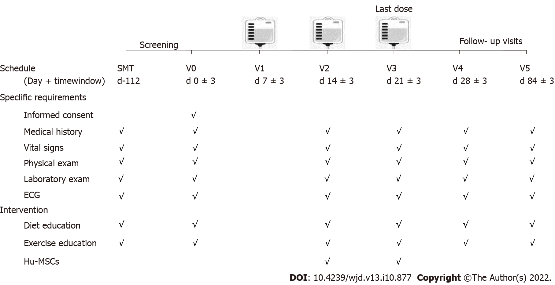
All patients were assessed again after 16 wk and were administered hUC-MSC infusions. The infusion was administered at a dosage of 1 × 10⁶ cells/kg per week for 3 wk. Considering the possible accidental episodes in the real-life that may interrupt the patients’ follow-up visits plan in due time, we set a flexible time range (± 3 d) at the patient’s discretion but which would not affect the safety and effectiveness of the study. This flexible schedule was structured for in-clinic evaluations to occur on day 14 ± 3, day 21 ± 3, day 28 ± 3 and day 84 ± 3 after the first dosage (Figure 1).
The effectiveness assessments were performed on day 14 ± 3, 21 ± 3, 28 ± 3, and 84 ± 3, including FBG, 2-h postprandial blood glucose, HbA1c, fasting CP (FCP), 2-h postprandial CP (P2CP), IR index [homeostatic model assessment for IR (HOMA-IR)] (by CP) = 1.5 + FCP ´ FBG/2800, islet β-cell function [HOMA of β-cell function (HOMA-β)] (by CP) = 0.27 ´ FCP/(FBG - 3.5), and hypoglycemic agent dosage. These dosages were adjusted by the treating physician according to standard clinical practice, based on blood glucose and A1c results.
Any adverse event after receiving the hUC-MSC infusions would be reported and recorded. Routine safety assessment was conducted according to the visit schedule, including blood routine examination, hepatic function test, electrocardiogram, chest radiography, and tumor-associated antigen test.
All statistical analyses were analyzed with SPSS® 25.0 software (IBM Corp, Armonk, NY, United States). Quantitative data were analyzed with the two-samples Wilcoxon test. Differences in proportions were analyzed by the two-tailed test. P < 0.05 was considered statistically significant.
A total of 16 T2DM patients were enrolled. The clinical characteristics are shown in Table 1.
| Characteristic | n = 16 |
| Age (yr) | 52.5 ± 7.91 |
| Male | 12 |
| Female | 4 |
| Duration of diabetes (yr) | 10.06 ± 5.74 |
| BMI (kg/m2) | 24.47 ± 2.76 |
| Glucose (mmol/L) | |
| FPG | 9.66 ± 2.65 |
| P2PG | 16.32 ± 4.64 |
| HbA1c (%) | 8.01 ± 0.63 |
| C peptide (pmol/L) | |
| FCP | 741.56 ± 464.50 |
| P2CP | 1596.70 ± 989.65 |
| HOMA-IR | 4.22 ± 1.91 |
| HOMA-β (%) | 35.01 ± 24.35 |
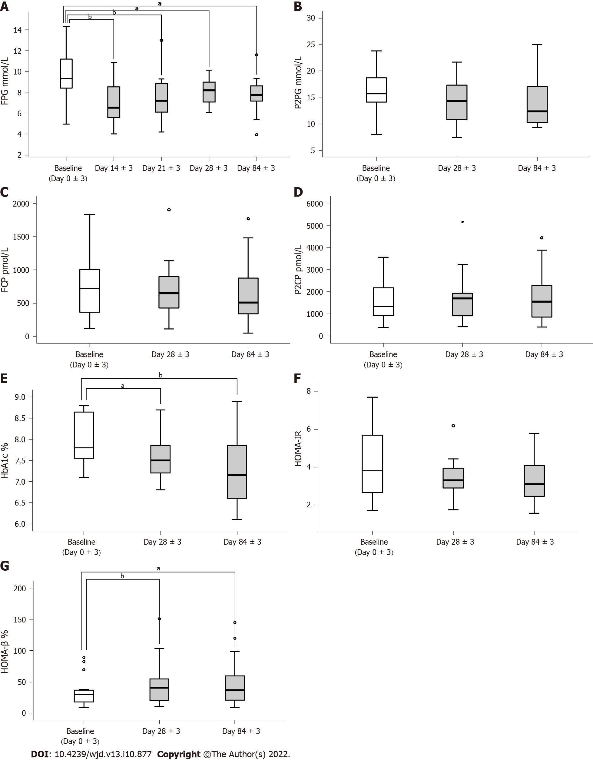
FPG was significantly reduced on day 14 ± 3 without any alteration of hypoglycemic drug dosage, achieving the lowest level during the entire intervention period [baseline: 9.3400 (8.3575, 11.7725), day 14 ± 3: 6.5200 (5.2200, 8.6900); P < 0.01]. The FBG had a sustained decrease during the follow-up visit period, with a reduction of hypoglycemic agents for all patients. HbA1c level was significantly reduced on day 84 ± 3 [baseline: 7.8000 (7.5250, 8.6750), day 84 ± 3: 7.150 (6.600, 7.925); P < 0.01] (Figure 2). There was no significant difference in postprandial blood glucose level in the 75-g oral glucose tolerance test without hypoglycemic agents.
The patients’ islet β-cell function was significantly improved on day 28 ± 3 [baseline: 29.90 (16.43, 37.40), day 28 ± 3: 40.97 (19.27, 56.36); P < 0.01]. Islet β-cell function (HOMA-β) improvement was stably sustained during the following intervention period, with a reduction in hypoglycemic agents in all patients. The HOMA-IR decreased but not to a level that was statistically significant (Figure 2).
After intravenous infusion of hUC-MSCs, the dosage of hypoglycemic agent was reduced in all patients on day 28 ± 3, of whom 12 (75%) had a decrement of 10%-50% and 4 (25%) had a decrement of 50%. On day 84 ± 3, the dosage was reduced in all patients, of whom 6 (50%) had a decrement of more than 50% and 1 (6.25%) discontinued the hypoglycemic agents (Figure 2).
Four patients had transient fever (5 times in total), which occurred within 24 h after the second or third infusion. One patient (2.08%) had asymptomatic nocturnal hypoglycemia after infusion on day 28 ± 3. It did not recur after reducing the dosage of insulin in the following period. No patient had acute diabetic complications during the intervention period.
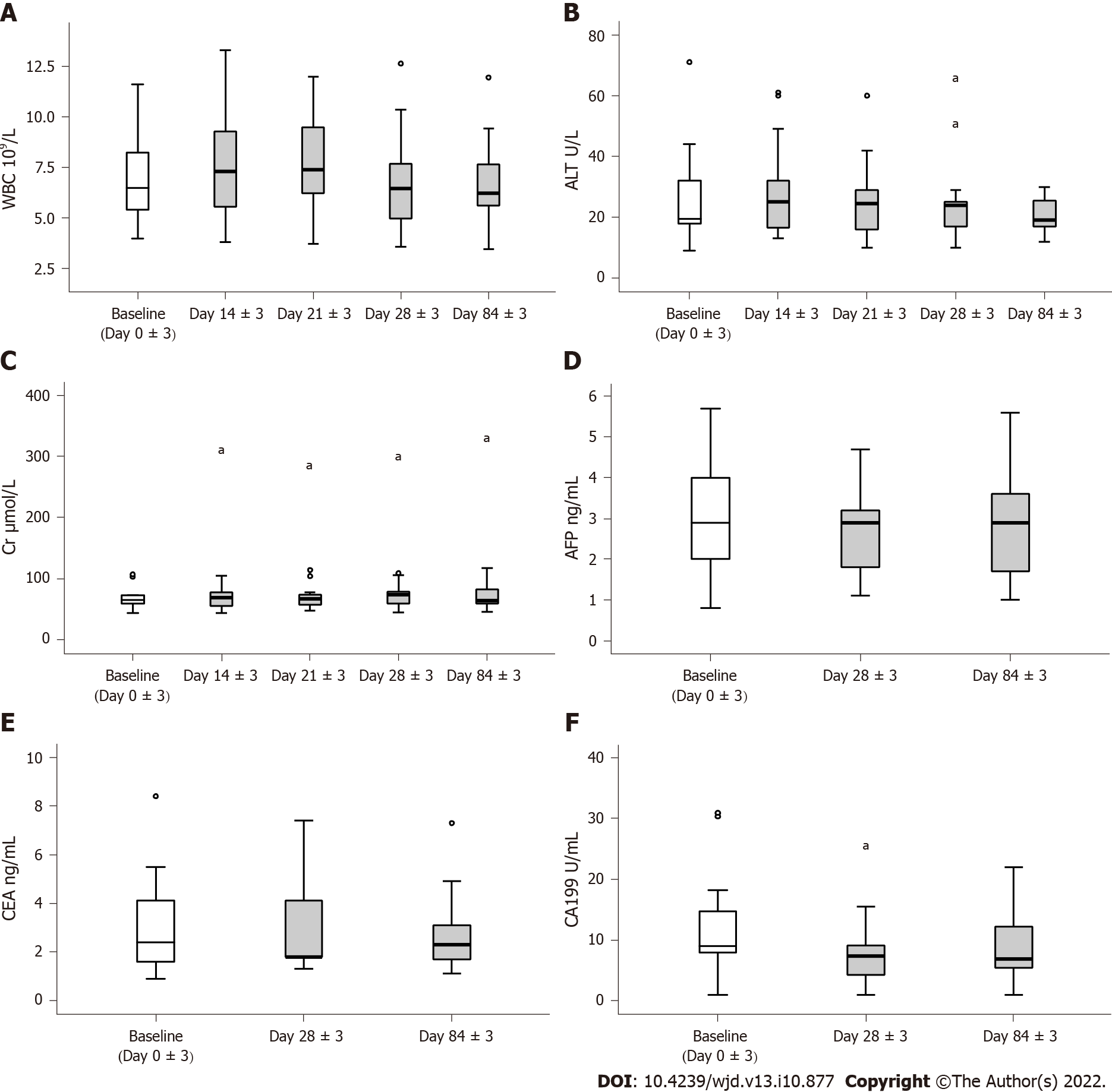
The leukocytes transiently increased significantly on day 14 ± 3 after the first dosage of hUC-MSCs, but there was no significant difference in leukocytes after the second dosage compared with baseline. The leukocyte level remained stable in the following period. There were no significant alterations in serum levels of alanine aminotransferase and creatinine (Figure 3).
After three doses of hUC-MSCs, carbohydrate antigen 199 and alpha fetoprotein decreased on day 28 ± 3. There was no significant alteration in carcinoembryonic antigen (Figure 3) or pancreatic autoantibody in the patients. All patients were negative for islet autoantibodies, with the exception of 1 who was positive for anti-islet cell antibody before and after the intervention.
Previous studies have demonstrated that hUC-MSCs are capable of decreasing the levels of FPG and HbA1c and reducing the dosage of hypoglycemic agents[13,22-24]. FBG was decreased and islet β-cell function was significantly improved after treatment with hUC-MSCs in our preliminary study in a diabetic rat model (data not shown). The purpose of this study was to evaluate the effectiveness and safety of hUC-MSC intravenous infusion in the short term for T2DM patients. The results demonstrated that hUC-MSCs could ameliorate hyperglycemia by decreasing FPG and HbA1c and reducing the dosage of hypoglycemic agents. It also improved islet β-cell function. Bell et al[25] observed the expression of pancreatic and duodenal homeobox 1 and islet cell differentiation gradually increased after hUC-MSC treatment of streptozotocin-treated NOD-SCID mice. MSC transplantation in streptozotocin-treated mice promoted the proliferation of endogenous islet cells and increased the amount of islet β-cells and insulin secretion[26]. Si et al[27] showed that the “increased” pancreatic islets and islet β-cells were not due to cell proliferation but to tissue repair and a decrease in apoptosis and damage in a rat model of T2D. Caplan et al[28] demonstrated that MSCs could ameliorate β-cell damage and restore islet β-cell function in a murine model in the early stage (7 d) of MSC treatment. Liu et al[12] reported that Wharton’s jelly-derived MSC transplantation increased the HOMA-β from 65.99 ± 23.49 % to 98.86 ± 43.91%. The clinical trial results of Hu et al[22] also showed that the HOMA-β was significantly increased 1 mo after hUC-MSC treatment and was maintained for 18 mo. The results of the current study were consistent with these findings, indicating that improvement in glycemia in T2DM patients after hUC-MSC treatment was related to the repair of islet β-cells.
IR plays a critical role in the development of T2DM. It has been reported that MSC treatment in the early stage improved IR in animal experiments[27]. Chen et al[24] showed that hUC-MSC treatment could increase the area under curve of the CP and decrease the HOMA-IR. Hu et al[22] showed that hUC-MSC treatment decreased the FCP and improved the HOMA-IR. However, the present study found no significant improvement of IR and no significant decrease in FCP and P2CP during the follow-up period. The islet β-cell function and IR state of patients in this study will be extensively followed for further analysis to elucidate the mechanisms underlying glycemia improvement.
Embryonic stem cells have the risk of teratoma formation, which limits their clinical application[29]. While the MSCs have been documented as having therapeutic efficacy for inflammation-related diseases, the concerns of possible tumorigenic effects are undeniable; although some studies have shown that MSCs do not undergo malignant transformation[30,31]. Guan et al[14] observed no immediate or delayed toxicity associated with MSC administration (within the followup period). In the present study, we observed no significant alterations in tumor-associated antigens (alpha-fetoprotein, carcinoembryonic antigen, carbohydrate antigen 199) within the follow-up period. Because the follow-up time was short, we plan to follow up the participants for 3 years for further observations of possible transplant complications.
As this was a preliminary exploratory study, our sample size was limited; we plan to recruit more participants and include a healthy control group in our future study to evaluate the clinical utility of this therapy for T2DM.
The results of our study suggest that hUC-MSC treatment can improve glycemia, restore islet βcell function, and safely reduce the dosage of hypoglycemic agents required by the patient. Thus, hUC-MSC treatment could be a novel therapy for T2DM.
Cellular therapies offer novel opportunities for the treatment of type 2 diabetes mellitus (T2DM) to improve the function of islet β-cells. However, the effectiveness and safety of human umbilical cord-mesenchymal stem cell (hUC-MSCs) in clinical application have not been fully assessed.
We conducted the present trial to explore the therapeutic effectiveness and mechanism of hUC-MSC infusion for treating T2DM.
We hypothesized that hUC-MSCs restore β-cell function by differentiating into β-cells. We conducted the present trial to treat T2DM with hUC-MSC infusion and evaluated the effectiveness and safety of hUC-MSC therapy.
Patients were enrolled and received 1 × 10⁶ cells/kg per week for 3 wk of intravenous hUC-MSC infusion. The effectiveness was assessed by fasting blood glucose, C-peptide, normal glycosylated hemoglobin A1c level (HbA1c), insulin resistance (IR) index (homeostasis model assessment of insulin resistance), islet β-cell function (homeostasis model assessment of β-cell function), and dosage of hypoglycemic agents, and the safety was evaluated by monitoring the occurrence of any adverse events.
During the entire intervention period, the fasting plasma glucose level and HbA1c were significantly reduced. The patients’ islet β-cell function was significantly improved, and the dosage of hypoglycemic agents was reduced in all patients without serious adverse events.
We hypothesize that hUC-MSCs restore β-cell function by differentiating into β-cells. Our study suggests that hUC-MSC treatment can improve glycemia, restore islet βcell function, and safely reduce the dosage of hypoglycemic agents.
Islet β-cell function and IR state of the patients in this study will be extensively followed for further analysis to elucidate the mechanisms underlying glycemia improvement.
The authors acknowledge the financial support from all of the funders. The authors also want to thank all patients and their families who consented to participate in the study.
Provenance and peer review: Unsolicited article; Externally peer reviewed.
Peer-review model: Single blind
Specialty type: Endocrinology and metabolism
Country/Territory of origin: China
Peer-review report’s scientific quality classification
Grade A (Excellent): 0
Grade B (Very good): 0
Grade C (Good): C, C
Grade D (Fair): 0
Grade E (Poor): 0
P-Reviewer: Bayoumi RAL, United Arab Emirates; Salim A, Pakistan S-Editor: Zhang H L-Editor: A P-Editor: Zhang H
| 1. | International Diabetes Federation. IDF diabetes atlas ninth. Dunia: Idf. 2019;9:5-9. [DOI] [Full Text] |
| 2. | Chinese Diabetes Society. Chinese clinical guidlines for T2DM (2017 Version). Zhonghuo Shiyong Nei Ke Za Zhi. 2018;38:292-344. [RCA] [DOI] [Full Text] [Cited by in Crossref: 8] [Cited by in RCA: 4] [Article Influence: 0.6] [Reference Citation Analysis (0)] |
| 3. | Cerf ME. Beta cell dysfunction and insulin resistance. Front Endocrinol (Lausanne). 2013;4:37. [RCA] [PubMed] [DOI] [Full Text] [Full Text (PDF)] [Cited by in Crossref: 404] [Cited by in RCA: 541] [Article Influence: 45.1] [Reference Citation Analysis (0)] |
| 4. | Bagust A, Beale S. Deteriorating beta-cell function in type 2 diabetes: a long-term model. QJM. 2003;96:281-288. [RCA] [PubMed] [DOI] [Full Text] [Cited by in Crossref: 77] [Cited by in RCA: 76] [Article Influence: 3.5] [Reference Citation Analysis (0)] |
| 5. | Levy J, Atkinson AB, Bell PM, McCance DR, Hadden DR. Beta-cell deterioration determines the onset and rate of progression of secondary dietary failure in type 2 diabetes mellitus: the 10-year follow-up of the Belfast Diet Study. Diabet Med. 1998;15:290-296. [RCA] [PubMed] [DOI] [Full Text] [Cited by in RCA: 4] [Reference Citation Analysis (0)] |
| 6. | U.K. prospective diabetes study 16. Overview of 6 years' therapy of type II diabetes: a progressive disease. U.K. Prospective Diabetes Study Group. Diabetes. 1995;44:1249-1258. [PubMed] |
| 7. | Gamble A, Pepper AR, Bruni A, Shapiro AMJ. The journey of islet cell transplantation and future development. Islets. 2018;10:80-94. [RCA] [PubMed] [DOI] [Full Text] [Cited by in Crossref: 124] [Cited by in RCA: 113] [Article Influence: 16.1] [Reference Citation Analysis (0)] |
| 8. | Zhang Y, Chen W, Feng B, Cao H. The Clinical Efficacy and Safety of Stem Cell Therapy for Diabetes Mellitus: A Systematic Review and Meta-Analysis. Aging Dis. 2020;11:141-153. [RCA] [PubMed] [DOI] [Full Text] [Full Text (PDF)] [Cited by in Crossref: 48] [Cited by in RCA: 48] [Article Influence: 9.6] [Reference Citation Analysis (0)] |
| 9. | He J, Kong D, Yang Z, Guo R, Amponsah AE, Feng B, Zhang X, Zhang W, Liu A, Ma J, O'Brien T, Cui H. Clinical efficacy on glycemic control and safety of mesenchymal stem cells in patients with diabetes mellitus: Systematic review and meta-analysis of RCT data. PLoS One. 2021;16:e0247662. [RCA] [PubMed] [DOI] [Full Text] [Full Text (PDF)] [Cited by in Crossref: 2] [Cited by in RCA: 5] [Article Influence: 1.3] [Reference Citation Analysis (1)] |
| 10. | Kamal MM, Kassem DH. Therapeutic Potential of Wharton's Jelly Mesenchymal Stem Cells for Diabetes: Achievements and Challenges. Front Cell Dev Biol. 2020;8:16. [RCA] [PubMed] [DOI] [Full Text] [Full Text (PDF)] [Cited by in Crossref: 55] [Cited by in RCA: 49] [Article Influence: 9.8] [Reference Citation Analysis (1)] |
| 11. | Pan XH, Song QQ, Dai JJ, Yao X, Wang JX, Pang RQ, He J, Li ZA, Sun XM, Ruan GP. Transplantation of bone marrow mesenchymal stem cells for the treatment of type 2 diabetes in a macaque model. Cells Tissues Organs. 2013;198:414-427. [RCA] [PubMed] [DOI] [Full Text] [Cited by in Crossref: 7] [Cited by in RCA: 12] [Article Influence: 1.1] [Reference Citation Analysis (0)] |
| 12. | Liu X, Zheng P, Wang X, Dai G, Cheng H, Zhang Z, Hua R, Niu X, Shi J, An Y. A preliminary evaluation of efficacy and safety of Wharton's jelly mesenchymal stem cell transplantation in patients with type 2 diabetes mellitus. Stem Cell Res Ther. 2014;5:57. [RCA] [PubMed] [DOI] [Full Text] [Full Text (PDF)] [Cited by in Crossref: 93] [Cited by in RCA: 121] [Article Influence: 11.0] [Reference Citation Analysis (0)] |
| 13. | Kong D, Zhuang X, Wang D, Qu H, Jiang Y, Li X, Wu W, Xiao J, Liu X, Liu J, Li A, Wang J, Dou A, Wang Y, Sun J, Lv H, Zhang G, Zhang X, Chen S, Ni Y, Zheng C. Umbilical cord mesenchymal stem cell transfusion ameliorated hyperglycemia in patients with type 2 diabetes mellitus. Clin Lab. 2014;60:1969-1976. [RCA] [PubMed] [DOI] [Full Text] [Cited by in Crossref: 37] [Cited by in RCA: 58] [Article Influence: 5.8] [Reference Citation Analysis (0)] |
| 14. | Guan LX, Guan H, Li HB, Ren CA, Liu L, Chu JJ, Dai LJ. Therapeutic efficacy of umbilical cord-derived mesenchymal stem cells in patients with type 2 diabetes. Exp Ther Med. 2015;9:1623-1630. [RCA] [PubMed] [DOI] [Full Text] [Cited by in Crossref: 44] [Cited by in RCA: 58] [Article Influence: 5.8] [Reference Citation Analysis (2)] |
| 15. | Tsai PJ, Wang HS, Lin GJ, Chou SC, Chu TH, Chuan WT, Lu YJ, Weng ZC, Su CH, Hsieh PS, Sytwu HK, Lin CH, Chen TH, Shyu JF. Undifferentiated Wharton's Jelly Mesenchymal Stem Cell Transplantation Induces Insulin-Producing Cell Differentiation and Suppression of T-Cell-Mediated Autoimmunity in Nonobese Diabetic Mice. Cell Transplant. 2015;24:1555-1570. [RCA] [PubMed] [DOI] [Full Text] [Cited by in Crossref: 26] [Cited by in RCA: 30] [Article Influence: 2.7] [Reference Citation Analysis (0)] |
| 16. | Wang G, Li Y, Wang Y, Dong Y, Wang FS, Ding Y, Kang Y, Xu X. Roles of the co-culture of human umbilical cord Wharton's jelly-derived mesenchymal stem cells with rat pancreatic cells in the treatment of rats with diabetes mellitus. Exp Ther Med. 2014;8:1389-1396. [RCA] [PubMed] [DOI] [Full Text] [Full Text (PDF)] [Cited by in Crossref: 7] [Cited by in RCA: 9] [Article Influence: 0.8] [Reference Citation Analysis (0)] |
| 17. | Ranjbaran H, Abediankenari S, Khalilian A, Rahmani Z, Momeninezhad Amiri M, Hosseini Khah Z. Differentiation of Wharton's Jelly Derived Mesenchymal Stem Cells into Insulin Producing Cells. Int J Hematol Oncol Stem Cell Res. 2018;12:220-229. [PubMed] |
| 18. | Domouky AM, Hegab AS, Al-Shahat A, Raafat N. Mesenchymal stem cells and differentiated insulin producing cells are new horizons for pancreatic regeneration in type I diabetes mellitus. Int J Biochem Cell Biol. 2017;87:77-85. [RCA] [PubMed] [DOI] [Full Text] [Cited by in Crossref: 16] [Cited by in RCA: 26] [Article Influence: 3.3] [Reference Citation Analysis (0)] |
| 19. | Lambert M. ADA releases revisions to recommendations for standards of medical care in diabetes. Am Fam Physician. 2012;85:514-515. [PubMed] |
| 20. | Gu J, Huang L, Zhang C, Wang Y, Zhang R, Tu Z, Wang H, Zhou X, Xiao Z, Liu Z, Hu X, Ke Z, Wang D, Liu L. Therapeutic evidence of umbilical cord-derived mesenchymal stem cell transplantation for cerebral palsy: a randomized, controlled trial. Stem Cell Res Ther. 2020;11:43. [RCA] [PubMed] [DOI] [Full Text] [Full Text (PDF)] [Cited by in Crossref: 61] [Cited by in RCA: 55] [Article Influence: 11.0] [Reference Citation Analysis (2)] |
| 21. | Dominici M, Le Blanc K, Mueller I, Slaper-Cortenbach I, Marini F, Krause D, Deans R, Keating A, Prockop Dj, Horwitz E. Minimal criteria for defining multipotent mesenchymal stromal cells. The International Society for Cellular Therapy position statement. Cytotherapy. 2006;8:315-317. [RCA] [PubMed] [DOI] [Full Text] [Cited by in Crossref: 11055] [Cited by in RCA: 12641] [Article Influence: 702.3] [Reference Citation Analysis (2)] |
| 22. | Hu J, Wang Y, Gong H, Yu C, Guo C, Wang F, Yan S, Xu H. Long term effect and safety of Wharton's jelly-derived mesenchymal stem cells on type 2 diabetes. Exp Ther Med. 2016;12:1857-1866. [RCA] [PubMed] [DOI] [Full Text] [Cited by in Crossref: 54] [Cited by in RCA: 64] [Article Influence: 7.1] [Reference Citation Analysis (0)] |
| 23. | Cai J, Wu Z, Xu X, Liao L, Chen J, Huang L, Wu W, Luo F, Wu C, Pugliese A, Pileggi A, Ricordi C, Tan J. Umbilical Cord Mesenchymal Stromal Cell With Autologous Bone Marrow Cell Transplantation in Established Type 1 Diabetes: A Pilot Randomized Controlled Open-Label Clinical Study to Assess Safety and Impact on Insulin Secretion. Diabetes Care. 2016;39:149-157. [RCA] [PubMed] [DOI] [Full Text] [Cited by in Crossref: 109] [Cited by in RCA: 139] [Article Influence: 15.4] [Reference Citation Analysis (0)] |
| 24. | Chen P, Huang Q, Xu XJ, Shao ZL, Huang LH, Yang XZ, Guo W, Li CM, Chen C. [The effect of liraglutide in combination with human umbilical cord mesenchymal stem cells treatment on glucose metabolism and β cell function in type 2 diabetes mellitus]. Zhonghua Nei Ke Za Zhi. 2016;55:349-354. [RCA] [PubMed] [DOI] [Full Text] [Cited by in RCA: 11] [Reference Citation Analysis (0)] |
| 25. | Bell GI, Meschino MT, Hughes-Large JM, Broughton HC, Xenocostas A, Hess DA. Combinatorial human progenitor cell transplantation optimizes islet regeneration through secretion of paracrine factors. Stem Cells Dev. 2012;21:1863-1876. [RCA] [PubMed] [DOI] [Full Text] [Cited by in Crossref: 36] [Cited by in RCA: 33] [Article Influence: 2.5] [Reference Citation Analysis (0)] |
| 26. | Karnieli O, Izhar-Prato Y, Bulvik S, Efrat S. Generation of insulin-producing cells from human bone marrow mesenchymal stem cells by genetic manipulation. Stem Cells. 2007;25:2837-2844. [RCA] [PubMed] [DOI] [Full Text] [Cited by in Crossref: 205] [Cited by in RCA: 198] [Article Influence: 11.0] [Reference Citation Analysis (0)] |
| 27. | Si Y, Zhao Y, Hao H, Liu J, Guo Y, Mu Y, Shen J, Cheng Y, Fu X, Han W. Infusion of mesenchymal stem cells ameliorates hyperglycemia in type 2 diabetic rats: identification of a novel role in improving insulin sensitivity. Diabetes. 2012;61:1616-1625. [RCA] [PubMed] [DOI] [Full Text] [Full Text (PDF)] [Cited by in Crossref: 182] [Cited by in RCA: 208] [Article Influence: 16.0] [Reference Citation Analysis (0)] |
| 28. | Caplan AI, Dennis JE. Mesenchymal stem cells as trophic mediators. J Cell Biochem. 2006;98:1076-1084. [RCA] [PubMed] [DOI] [Full Text] [Cited by in Crossref: 2126] [Cited by in RCA: 2222] [Article Influence: 116.9] [Reference Citation Analysis (0)] |
| 29. | Thomson JA, Itskovitz-Eldor J, Shapiro SS, Waknitz MA, Swiergiel JJ, Marshall VS, Jones JM. Embryonic stem cell lines derived from human blastocysts. Science. 1998;282:1145-1147. [RCA] [PubMed] [DOI] [Full Text] [Cited by in Crossref: 11399] [Cited by in RCA: 10416] [Article Influence: 385.8] [Reference Citation Analysis (0)] |
| 30. | Wang Y, Zhang Z, Chi Y, Zhang Q, Xu F, Yang Z, Meng L, Yang S, Yan S, Mao A, Zhang J, Yang Y, Wang S, Cui J, Liang L, Ji Y, Han ZB, Fang X, Han ZC. Long-term cultured mesenchymal stem cells frequently develop genomic mutations but do not undergo malignant transformation. Cell Death Dis. 2013;4:e950. [RCA] [PubMed] [DOI] [Full Text] [Full Text (PDF)] [Cited by in Crossref: 107] [Cited by in RCA: 123] [Article Influence: 10.3] [Reference Citation Analysis (0)] |
| 31. | Fu Y, Li J, Li M, Xu J, Rong Z, Ren F, Wang Y, Sheng J, Chang Z. Umbilical Cord Mesenchymal Stem Cells Ameliorate Inflammation-Related Tumorigenesis via Modulating Macrophages. Stem Cells Int. 2022;2022:1617229. [RCA] [PubMed] [DOI] [Full Text] [Full Text (PDF)] [Cited by in Crossref: 2] [Cited by in RCA: 5] [Article Influence: 1.7] [Reference Citation Analysis (0)] |


