Copyright
©The Author(s) 2015.
World J Diabetes. Feb 15, 2015; 6(1): 37-53
Published online Feb 15, 2015. doi: 10.4239/wjd.v6.i1.37
Published online Feb 15, 2015. doi: 10.4239/wjd.v6.i1.37
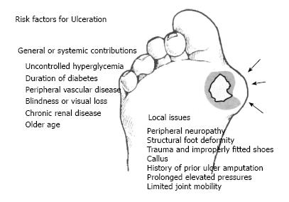
Figure 1 The risk factors for diabetic foot ulcer.
Ulcers may be distinguished by general or systemic considerations vs those localized to the foot and its pathology. (Data adapted from Frykberg et al[18]).
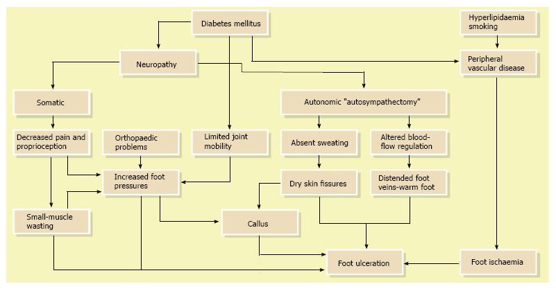
Figure 2 Etiology of diabetic foot ulcer.
(Data adapted from Boulton et al[17]).
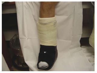
Figure 3 Total contact cast for patients with diabetic foot ulcer.
(Data adapted from Armstrong et al[82]).
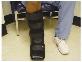
Figure 4 Removable cast walker (DH Walker) for patients with diabetic foot ulcer.
(Data adapted from Rathur et al[86]).
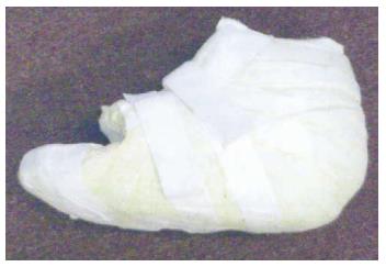
Figure 6 Scotch-cast boot for off-loading pressure from the foot of a diabetic patient with foot ulcer.
(Data adapted from Armstrong et al[82]).
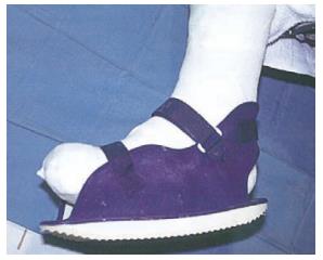
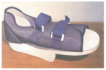
Figure 7 Half shoe for off-loading pressure from the foot of a diabetic patient with foot ulcer.
(Data adapted from Armstrong et al[82]).
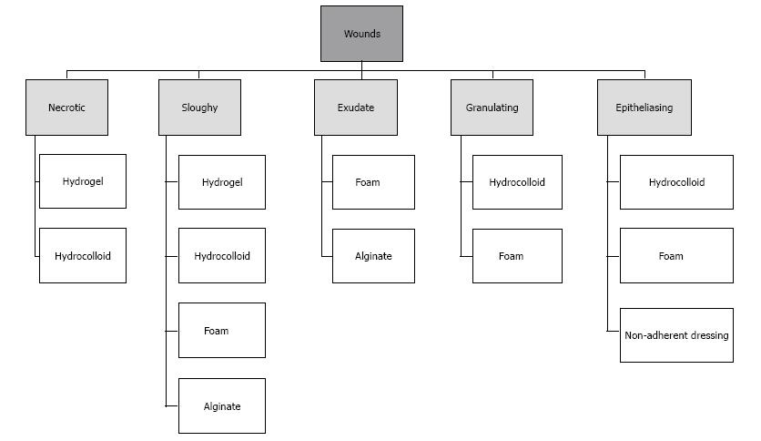
Figure 8 Classification of the different advanced dressing types usually used in diabetic foot ulcer treatment.
(Data adapted from Moura et al[95]).
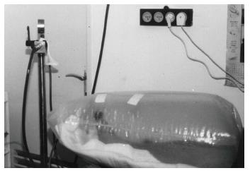
Figure 9 The polyethylene hyperbaric chamber.
Oxygen in a concentration of 100% was pumped into the bag through a regular car wheel valve. The open end of the bag was sealed by an elastic bandage to the leg above the knee. Oxygen was allowed to leak around the bandage, and the pressure in the chamber was kept to between 20 and 30 mmHg (1.02-1.03 atm) above atmospheric pressure. (Data adapted from Landau[127]).
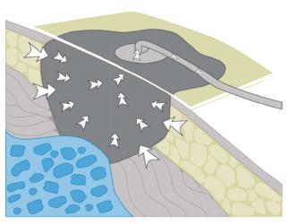
Figure 10 Schematic drawing of the negative pressure wound treatment.
(Data adapted from Vikatmaa et al[147]).
- Citation: Yazdanpanah L, Nasiri M, Adarvishi S. Literature review on the management of diabetic foot ulcer. World J Diabetes 2015; 6(1): 37-53
- URL: https://www.wjgnet.com/1948-9358/full/v6/i1/37.htm
- DOI: https://dx.doi.org/10.4239/wjd.v6.i1.37


