Copyright
©The Author(s) 2015.
World J Diabetes. Feb 15, 2015; 6(1): 151-166
Published online Feb 15, 2015. doi: 10.4239/wjd.v6.i1.151
Published online Feb 15, 2015. doi: 10.4239/wjd.v6.i1.151
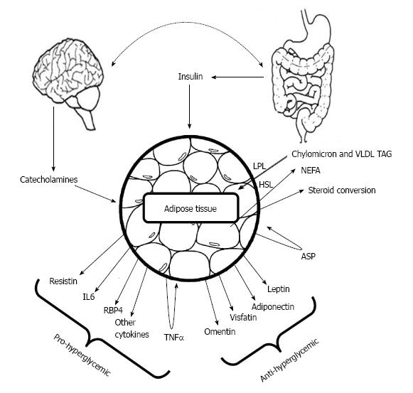
Figure 1 Adipocyte-derived proteins with anti-diabetic actions include leptin, adiponectin, omentin and visfatin; other factors tend to raise blood glucose including resistin, Tumor necrosis factor-α and Retinol-binding protein 4.
(Adapted from Mohamed-Ali et al[31] and Rosen et al[32]). LPL: Lipoprotein lipase; HSL: Hormone-sensitive lipase; NEFA: Non-esterified fatty acids; ASP: Acylation stimulating protein; TAG: Triacylglycerol; TNF-α: Tumor necrosis factor α; RBP4: Retinol-binding protein; IL6: Interleukin 6.
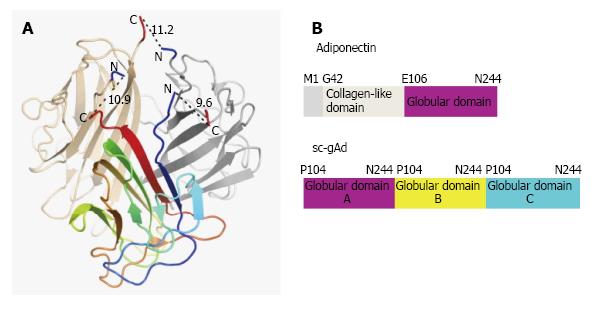
Figure 2 Structure of single-chain globular domain adiponectin (sc-gAd).
A: Base region of mouse gAd structure where blue arrow determines the N terminus and red arrow determines the C terminus; B: Domain organization of human adiponectin and the sc-gAd, where there are three domains A, B and C respectively. (Adapted from Min et al[30]).
Figure 3 Proposed structure of adiponectin receptors (Adapted from Kadowaki et al[36]).
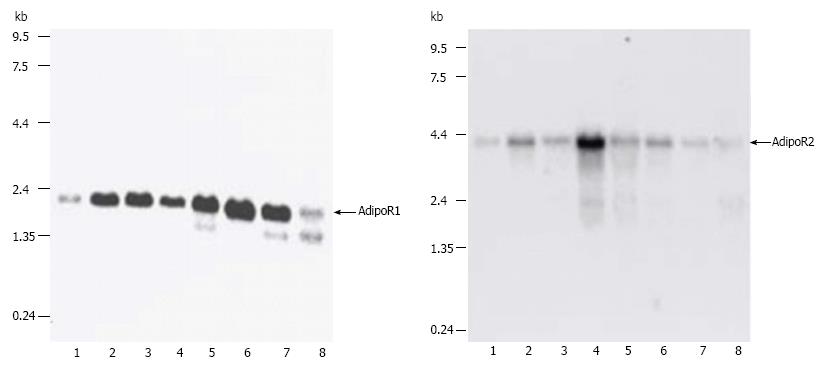
Figure 4 Northern blot analysis of AdipoR1 (top panel) and AdipoR2 (bottom panel) mRNA in mouse tissues (lanes: 1, brain; 2, heart; 3, kidney; 4, liver; 5, lung; 6, skeletal muscle; 7, spleen; 8, testis)[35].
AdipoR: Adiponectin receptors.
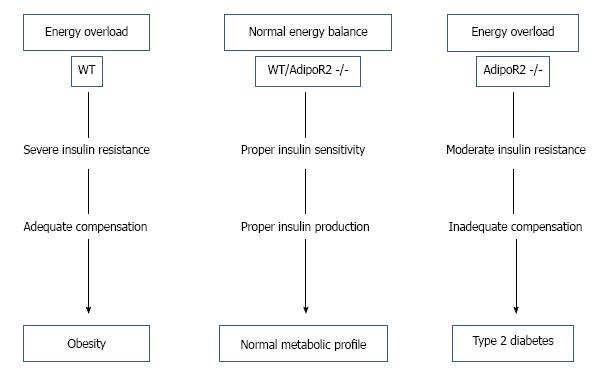
Figure 5 Diagram depicting the metabolic profile of wild type and AdipoR2 -/- mice (Adapted from Liu et al[38]).
WT: Wild type; AdipoR 2: Adiponectin receptors 2.
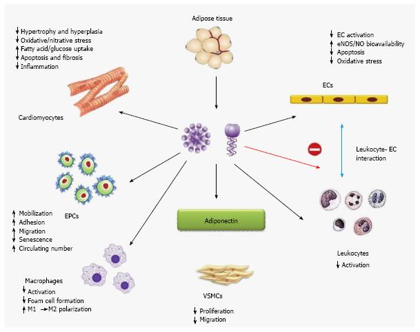
Figure 6 Actions of adiponectin in different cell line (Adapted from Xu et al[42]).
EC: Endothelial cell; EPCs: Endothelial progenitor cells; VSMC: Vascular smooth muscles; eNOS: Endothelial nitric oxide synthase.
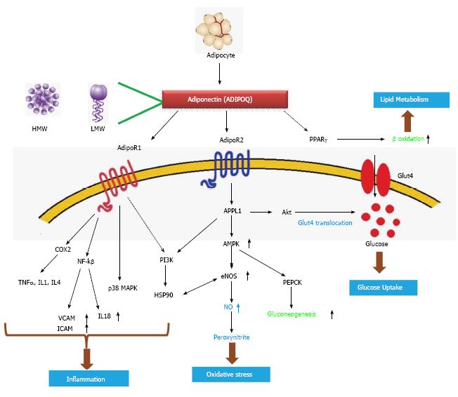
Figure 7 A proposed models of adiponectin metabolic pathway and associated genes.
HMW: High molecular weight; LMW: Low molecular weight AdipoR1: Adiponectin receptor 1; PPARγ: Peroxisome proliferator-activated receptor gamma; Glut4: Glucose transporter type 4; APPL1: Adaptor protein, phosphotyrosine interaction, pH domain and leucine zipper containing 1; Akt: Protein kinase B; COX2: Cyclooxygenase 2; AMPK: Adenosine monophosphate-activated protein kinase; PI3K: Phosphoinositide 3-kinase; NF-kβ: Nuclear factor kappa-light-chain-enhancer of activated B cells; TNFα: Tumor necrosis factor alpha; IL: Interleukin; p38 MAPK: p38 mitogen-activated protein kinase; HSP90: Heat shock protein 90; eNOS: Endothelial nitric oxide synthase; PEPCK: Phosphoenolpyruvate carboxykinase; NO: Nitric oxide; VCAM: Vascular cell adhesion protein; ICAM: Intercellular adhesion molecule.
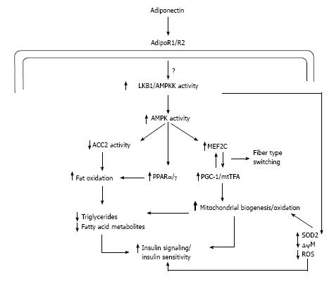
Figure 8 Hypothetical scheme of adiponectin signaling and the regulation of mitochondrial function in skeletal muscle (Adapted from Civitarese et al[59]).
AdipoR1: Adiponectin receptor 1; PPARγ: Peroxisome proliferator-activated receptor gamma; LKB1: Liver kinase B1; AMPKK: Adenosine monophosphate-activated protein kinase kinase; AMPK: Adenosine monophosphate-activated protein kinase; ACC2: Acetyl-CoA carboxylase 2; MEF2C: Myocyte-specific enhancer factor 2C; PPARα: Peroxisome proliferator-activated receptor alpha; PGC-1: Peroxisome proliferator-activated receptor gamma coactivator 1; mtTFA: Mitochondrial transcription factor A; SOD2: Superoxide dismutase 2; ΔψM: Mitochondrial membrane potential; ROS: Reactive oxygen species.
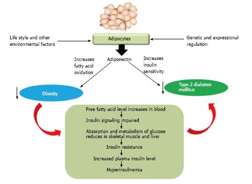
Figure 9 Hypothetical model showing the interrelation between adiponectin, obesity and type 2 diabetes mellitus.
- Citation: Ghoshal K, Bhattacharyya M. Adiponectin: Probe of the molecular paradigm associating diabetes and obesity. World J Diabetes 2015; 6(1): 151-166
- URL: https://www.wjgnet.com/1948-9358/full/v6/i1/151.htm
- DOI: https://dx.doi.org/10.4239/wjd.v6.i1.151


