Copyright
©2014 Baishideng Publishing Group Inc.
World J Diabetes. Oct 15, 2014; 5(5): 666-677
Published online Oct 15, 2014. doi: 10.4239/wjd.v5.i5.666
Published online Oct 15, 2014. doi: 10.4239/wjd.v5.i5.666
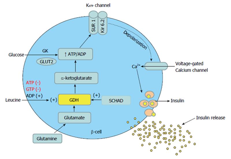
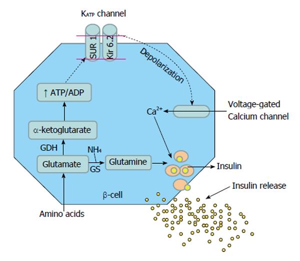
Figure 1 Glucose and protein mediated insulin secretion in the beta cell of pancreas.
GDH: Glutamate dehydrogenase; SCHAD: Short-chain 3-hydroxyacyl- CoA dehydrogenase; GK: Glucokinase; SUR: Sulfonylurea receptor; Kir 6.2: Potassium channel inwardly rectifying; GLUT2: Glucose transporter 2.
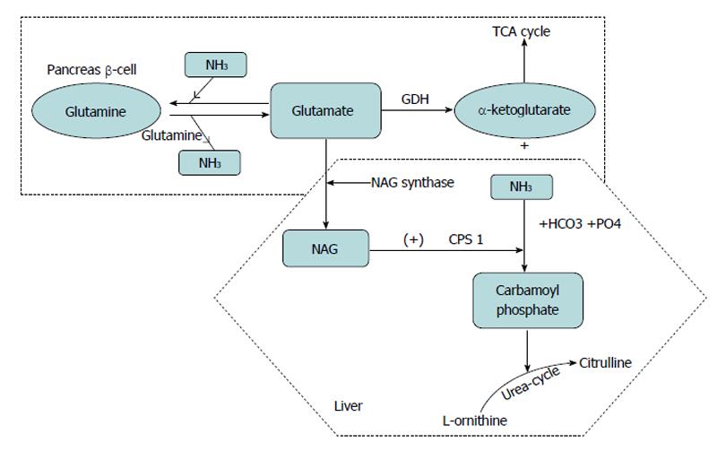
Figure 2 Glutamate metabolism.
Oxidation of glutamate by glutamate dehydrogenase liberates free ammonia (NH3) and alpha ketoglutarate, which enters tricarboxylic acid cycle cycle and generates ATP. In the liver glutamate also generates N-acetylglutamate (NAG), which in turn allosterically activates carbomyl phosphate synthetase (CPS) to regulate ammonia detoxification into urea. Glutamine provides a substrate for ammonia buffering, by adding ammonia to glutamate to form glutamine. TCA: Tricarboxylic acid cycle; GDH: Glutamate dehydrogenase.
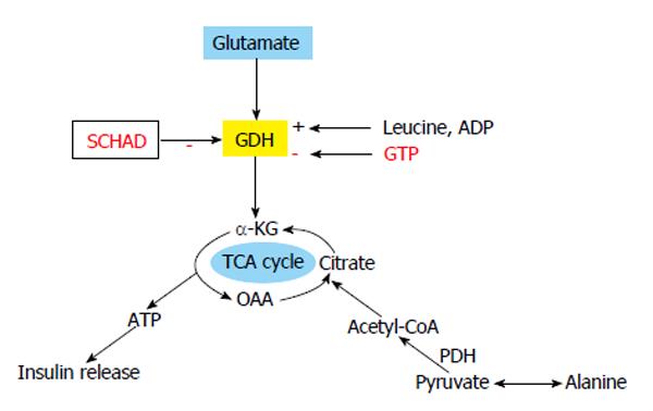
Figure 3 Glutamate and alanine as insulin secretagogues.
Protein induced hyperinsulinaemic hypoglycaemia due to loss of function mutation in HADH gene (SCHAD). Alanine is deaminated to pyruvate and pyruvate dehydrogenase (PDH) converts it to acetyl CoA, which can enter TCA cycle to generate ATP for closing KATP channel. TCA: Tricarboxylic acid cycle; α-KG: Alpha ketoglutarate; GDH: Glutamate dehydrogenase; OAA: Oxaloacetic acid.
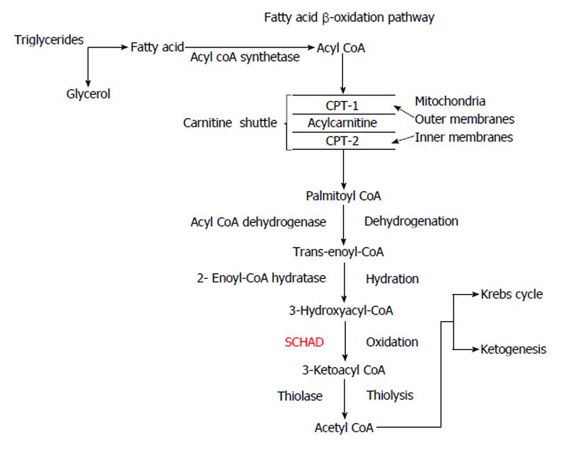
Figure 4 β-oxidation of fatty acids.
Acyl-CoA is converted to acetyl-CoA through dehydrogenation, hydration, oxidation and thiolysis. Acetyl-CoA can enter the Krebs cycle or can lead to ketogenesis. CPT-1: Carnitine palmitoyltransferase-1; SCHAD: 3-hydroxyacyl CoA dehydrogenase.
Figure 5 Protein Induced Hypoglycaemia due to defects in KATP channel genes.
GDH: Glutamate dehydrogenase; GK: Glucokinase.
- Citation: Chandran S, Yap F, Hussain K. Molecular mechanisms of protein induced hyperinsulinaemic hypoglycaemia. World J Diabetes 2014; 5(5): 666-677
- URL: https://www.wjgnet.com/1948-9358/full/v5/i5/666.htm
- DOI: https://dx.doi.org/10.4239/wjd.v5.i5.666


