Copyright
©2014 Baishideng Publishing Group Inc.
World J Diabetes. Jun 15, 2014; 5(3): 244-257
Published online Jun 15, 2014. doi: 10.4239/wjd.v5.i3.244
Published online Jun 15, 2014. doi: 10.4239/wjd.v5.i3.244
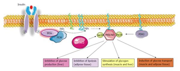
Figure 1 Insulin signaling pathway.
Insulin binds with insulin receptor (IR) that activates the IR substrates (IRSs), the phosphoinositide-3-kinase (PI3K), and protein kinase B/Akt (PKBAkt). Activated PKB/Akt mediates insulin metabolic effects and regulates nutrients homeostasis. PIP2: Phosphorylate phosphatidylinositol, 4, 5-bisphosphate; PDK1: 3-phosphoinositide-dependent protein kinase 1; mTORC2: Mammalian target of rapamycin complex 2.
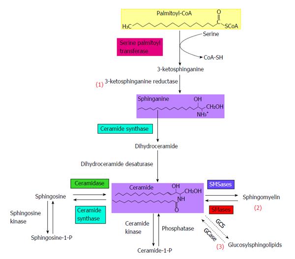
Figure 2 Sphingolipid metabolism.
Ceramide can either be newly synthesized in de novo ceramide synthesis pathway (1), or it can be the product of complex sphingolipids degradation, including sphingomyelin hydrolysis (2). The degradation of glycosylsphingolipids constitutes the salvage pathway (3). GCase: Glucosyl ceramidase; GCS: Glucosylceramide synthase; SMases: Sphingomyelinases; SMSases: Sphingomyelin synthases.
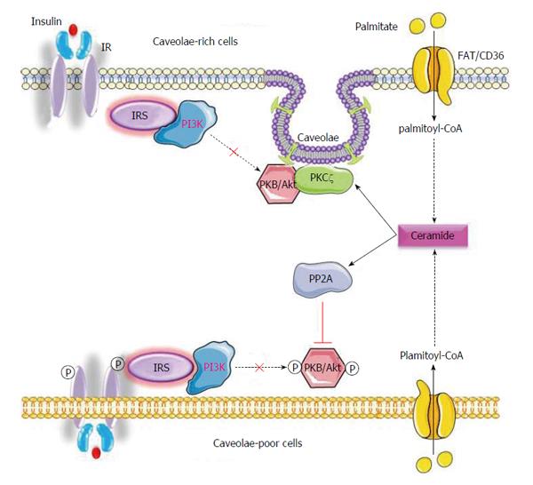
Figure 3 Ceramide inhibits insulin-induced activation of protein kinase B via two distinct mechanisms.
Ceramide inhibits insulin-activation of protein kinase B (PKB/Akt) either by activating atypical PKC (PKCζ) or by stimulating the phosphatase PP2A. Activation of either mechanism depends on plasma membrane enrichment with caveolae: (1) Ceramide-activated PKCζ phosphorylates the PH domain of PKB/Akt on Thr/Ser34, changing the recognition site of PKB/Akt, and disabling its activation by PIP3; (2) PP2A dephosphorylates PKB/Akt, inhibiting its kinase activity. IR: Insulin receptor; IRS: IR substrates; PI3K: Phosphoinositide-3-kinase; PP2A: Protein phosphatase 2A.
- Citation: Hage Hassan R, Bourron O, Hajduch E. Defect of insulin signal in peripheral tissues: Important role of ceramide. World J Diabetes 2014; 5(3): 244-257
- URL: https://www.wjgnet.com/1948-9358/full/v5/i3/244.htm
- DOI: https://dx.doi.org/10.4239/wjd.v5.i3.244


