Copyright
©2013 Baishideng Publishing Group Co.
World J Diabetes. Dec 15, 2013; 4(6): 310-318
Published online Dec 15, 2013. doi: 10.4239/wjd.v4.i6.310
Published online Dec 15, 2013. doi: 10.4239/wjd.v4.i6.310
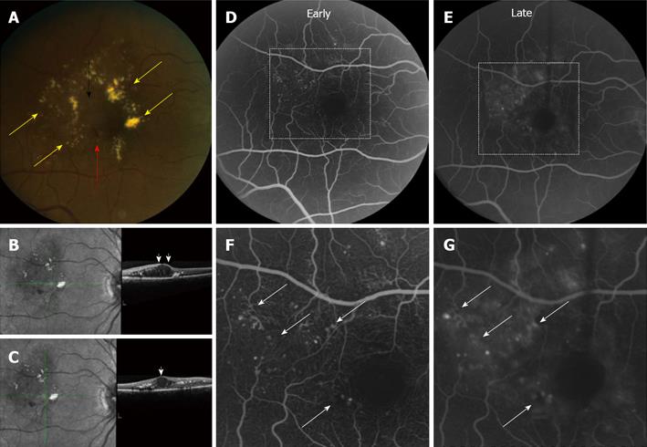
Figure 1 Fundus photo, optical coherence tomography, and fluorescein angiography of a patient with diabetic macular edema.
A: Fundus photo demonstrating classic presentation of diabetic macular edema with lipid exudate (yellow arrows), retinal thickening (black arrow), and intraretinal hemorrhages (red arrow); B, C: Horizontal (above) and vertical (below) high-resolution line scan demonstrating the presence of intraretinal cysts (white arrowheads) in the inner retina; D, E: Fluorescein angiographic images demonstrating focal leakage arising from microaneurysms (white arrows); F, G: Diffuse leakage arising from the walls of capillaries.
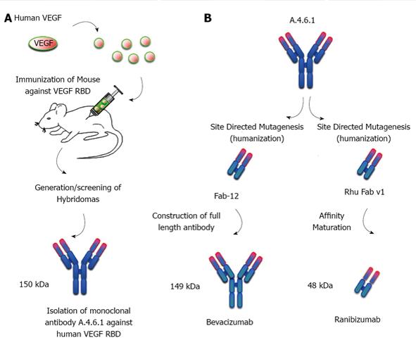
Figure 2 Development of humanized neutralizing monoclonal antibodies against vascular endothelial growth factor.
A: A polypeptide from the vascular endothelial growth factor (VEGF) receptor-binding domain (RBD) was used to immunize mice. The spleen from the mice was isolated and lymphocytes were cultured with immortal myeloma cell lines. Fusion of the cell lines results in the generation of hybridomas. Supernatants from the hybridoma cultures are tested for antibodies that bind specifically and with high affinity to VEGF. This resulted in the identification of the parental mouse monoclonal antibody, A.4.6.1; B: Site directed mutagenesis was then used to “humanize” the Fab (epitope-binding) fragment of A.4.6.1. Fab-12 was used to construct the full-length antibody, bevacizumab (left). Rhu Fab v1 was further affinity matured to produce the Fab fragment, ranibizumab (right).
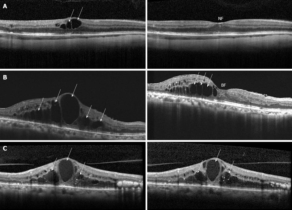
Figure 3 High resolution optical coherence tomography demonstrating different responses to treatment of diabetic macular edema patients with ranibizumab.
A: Modest cystoid macular edema (CME) with few inner retinal cysts (white arrows) and loss of the foveal contour (left) which completely resolved with return of a normal foveal contour (NF) and excellent vision one month after a single injection of ranibizumab (right); B: Massive CME with numerous inner and outer retinal cysts (white arrows) with complete loss of the foveal contour (left) which partially resolves resulting in a blunted but improved foveal contour (BF) and a significant improvement in vision following treatment with ranibizumab (right); C: Massive CME with numerous inner and outer retinal cysts (white arrows) with complete loss of the foveal contour (left) which does not respond despite repeated treatment with ranibizumab (right). The last response is uncommon; this patient was ultimately treated with intraocular steroids, and did have a sustained improvement of the edema and a modest improvement in vision.
- Citation: Krispel C, Rodrigues M, Xin X, Sodhi A. Ranibizumab in diabetic macular edema. World J Diabetes 2013; 4(6): 310-318
- URL: https://www.wjgnet.com/1948-9358/full/v4/i6/310.htm
- DOI: https://dx.doi.org/10.4239/wjd.v4.i6.310


