Copyright
©2013 Baishideng Publishing Group Co.
World J Diabetes. Dec 15, 2013; 4(6): 282-289
Published online Dec 15, 2013. doi: 10.4239/wjd.v4.i6.282
Published online Dec 15, 2013. doi: 10.4239/wjd.v4.i6.282
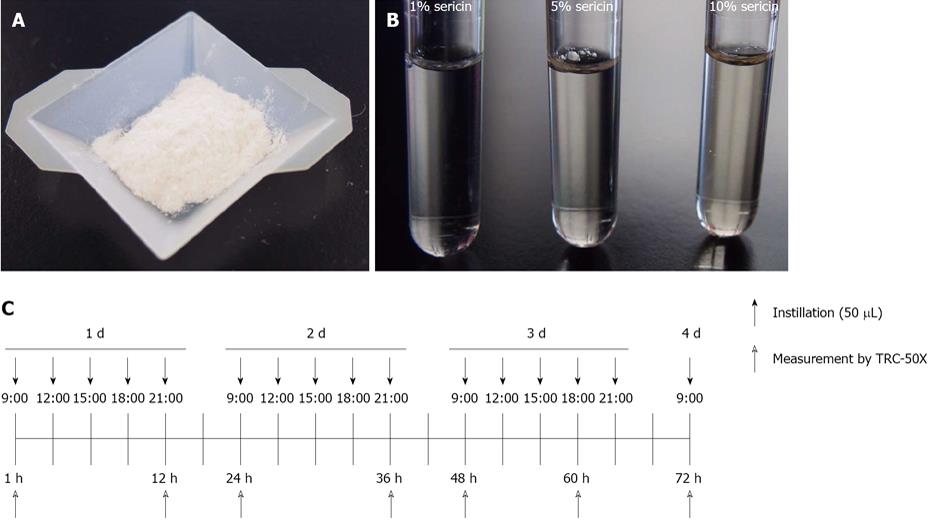
Figure 1 Picture of sericin and protocol in this study.
A: Picture of sericin; B: Picture of sericin solution. The sericin solutions used in this study were prepared by adding sericin to saline (pH 6.5-7.5); C: Protocol for instillation of sericin. Saline, sericin or hyaluronic acid solutions were instilled into the eyes of rats five times a day.
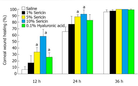
Figure 2 Corneal images of Wistar rats with or without the instillation of sericin solutions.
The corneal epithelium was removed with a BD Micro-Sharp™ and the resulting corneal wounds were dyed with 1% fluorescein solution. Saline, sericin or hyaluronic acid solutions were instilled into the eyes of rats five times a day. aP < 0.05 vs saline-instilled rat.
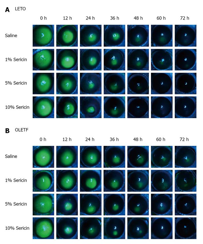
Figure 3 Corneal images.
A: Long-Evans Tokushima Otsuka (LETO) rats; B: Otsuka Long-Evans Tokushima Fatty (OLETF) rats with or without the instillation of sericin solutions. The photograph was reported in reference 39. The corneal epithelium was removed with a BD Micro-Sharp™ and the resulting corneal wounds were dyed with 1% fluorescein solution. Saline or sericin solutions were instilled into the eyes of rats five times a day.
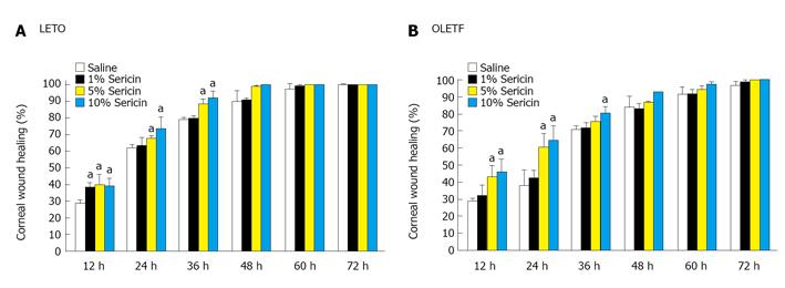
Figure 4 Effect of sericin solutions on corneal wound healing.
A: Long-Evans Tokushima Otsuka (LETO) rats; B: Otsuka Long-Evans Tokushima Fatty (OLETF) rat eyes. The data were reported in reference 39. Saline or sericin solutions were instilled into the eyes of rats five times a day. The data are presented as mean ± SE of 3-5 independent rat corneas. aP < 0.05 vs saline-instilled rat.
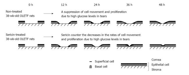
Figure 5 The function of cell migration and proliferation in corneal wound healing in 38 wk old Otsuka Long-Evans Tokushima Fatty rats with or without the instillation of sericin solutions.
The movement of superficial cells shows cell migration and the number of basal cells represents cell proliferation. OLETF: Otsuka Long-Evans Tokushima Fatty.
Figure 6 Effect of sericin on the adhesion (A) and growth (B) of human cornea epithelial cell line.
The data were reported in reference 51. Human cornea epithelial cell line (HCE-T) cells were cultured in Dulbecco’s modified Eagle’s medium/Ham’s F12 containing 5% (v/v) heat-inactivated fetal bovine serum, 0.1 mg/mL streptomycin and 1000 IU/mL penicillin. Cell growth was calculated by TetraColor One. The amount of cell adhesion and growth were represented by the following equation: cell adhesion or growth (%) = Abssericin treatment/Abscontrol× 100. The data are presented as mean ± SE of 5-25 experiments. aP < 0.05 vs control HCE-T cells.
- Citation: Nagai N, Ito Y. Therapeutic effects of sericin on diabetic keratopathy in Otsuka Long-Evans Tokushima Fatty rats. World J Diabetes 2013; 4(6): 282-289
- URL: https://www.wjgnet.com/1948-9358/full/v4/i6/282.htm
- DOI: https://dx.doi.org/10.4239/wjd.v4.i6.282


