Copyright
©The Author(s) 2024.
World J Diabetes. Jul 15, 2024; 15(7): 1499-1508
Published online Jul 15, 2024. doi: 10.4239/wjd.v15.i7.1499
Published online Jul 15, 2024. doi: 10.4239/wjd.v15.i7.1499
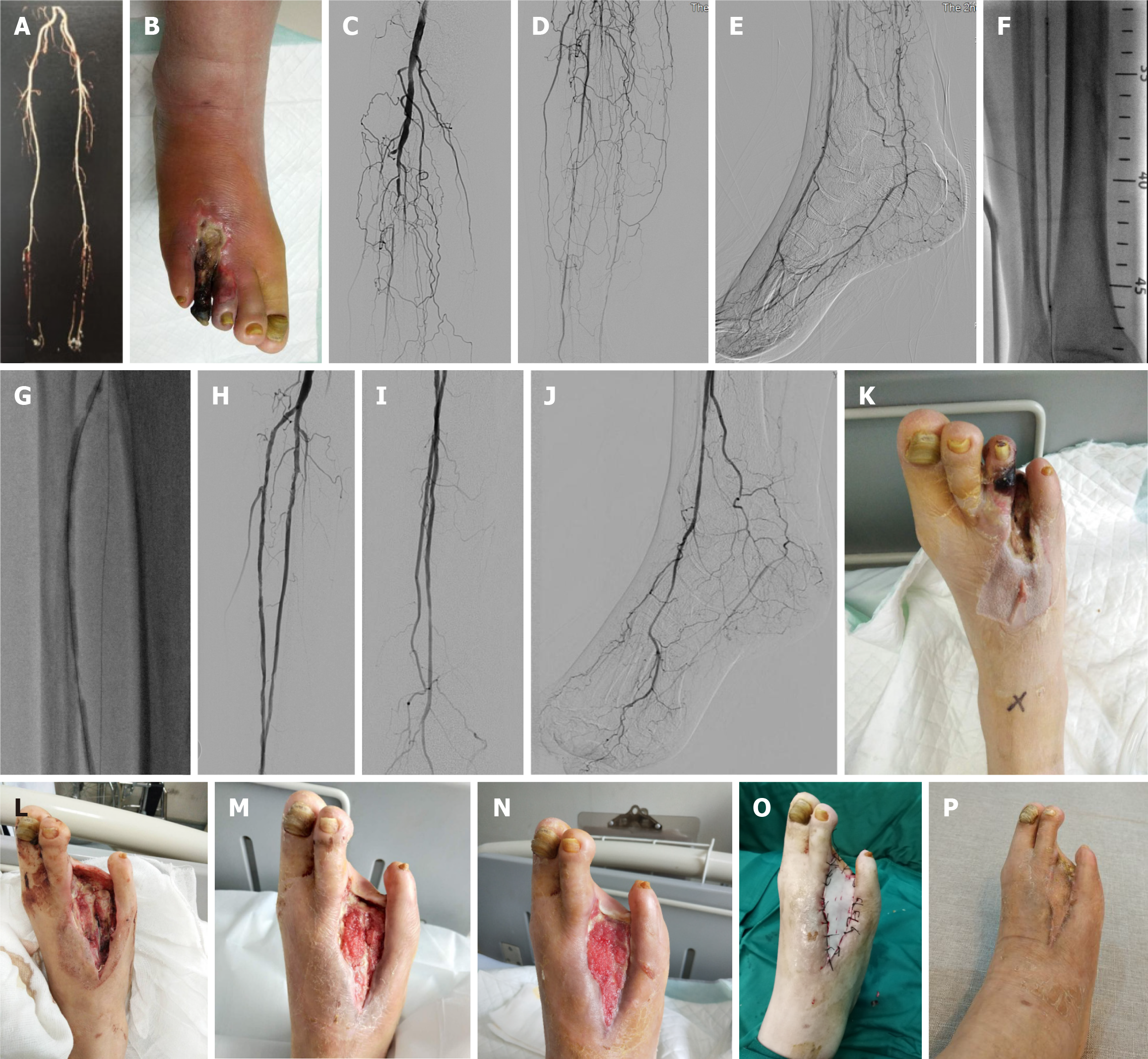
Figure 1 Surgery pictures of infrapopliteal artery occlusion case.
A: Lower extremity computerized tomography angiography suggests infrapopliteal artery occlusion; B: Gangrene of the fourth toe with infection; C-E: Long-segment occlusions of the anterior tibial artery, posterior tibial artery and peroneal artery; distal dorsal pedis artery and posterior tibial artery were demonstrated through lateral branches; F and G: The anterior tibial artery and peroneal artery were dilated by balloon angioplasty (BA) during the operation; H-J: After BA, the anterior tibial artery and peroneal artery were unobstructed, and the communication between lateral plantar artery and peroneal artery was developed; K and L: After revascularization, local debridement, removal of necrotic tissue and the 3rd and 4th phalanx, and vacuum-assisted closure (VAC) aspiration were performed; M: The wound surface two weeks after VAC; N: The wound granulation tissue grew well three weeks after VAC and reached the conditions for skin grafting; O: Skin grafting with split-thickness skin graft from the inner leg; P: Wound repair two months after skin grafting.
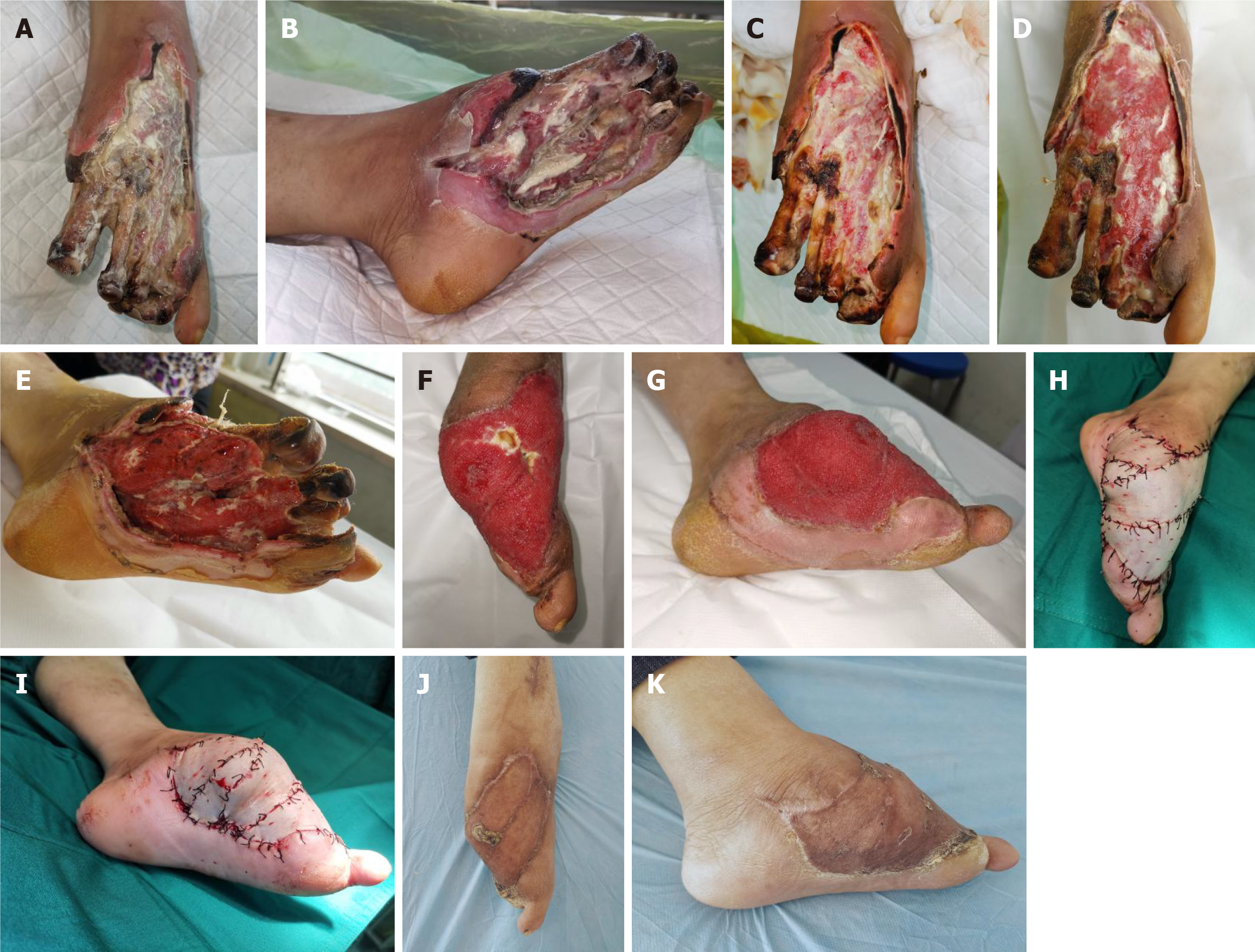
Figure 2 Surgery pictures of foot infection case.
A and B: Manifestations of foot infection after the patient's self-trimming of the plantar calluses and subsequent debridement in another hospital; C-E: After two weeks of nibbling debridement, the local granulation tissue of the patient began to grow; F and G: The granulation tissue grew well after two weeks of intermittent vacuum-assisted closure (VAC) after revascularization; H and I: Skin grafting was performed with split-thickness skin flaps from both thighs, and postoperative VAC was applied for one week; J and K: The appearance of the patient's foot one month after skin grafting.
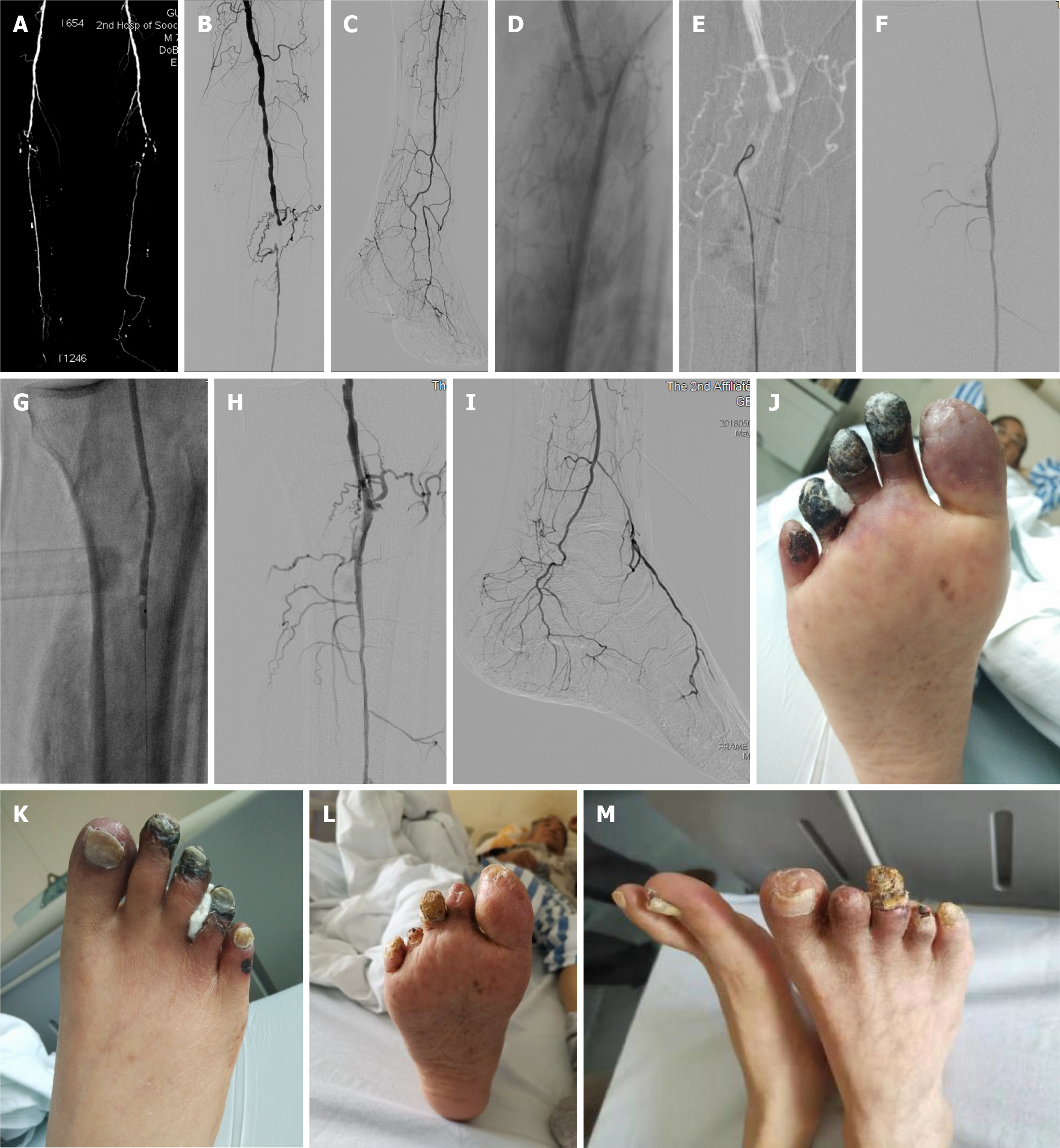
Figure 3 Surgery pictures of a diabetic foot complicated with uremia.
A: Lower limb computerized tomography angiography of a diabetic foot complicated with uremia showed proximal occlusion of the peroneal artery, the only outflow tract below the knee, obvious arterial calcification; B-D: Angiography showed that the proximal peroneal artery was occluded, and the guidewire could not pass through the lesion due to severe calcification; E-G: Retrograde puncture of the distal peroneal artery was performed, and balloon angioplasty (BA) was carried out after the orbit was established by connecting the guidewire with the proximal catheter; H and I: The blood flow was obviously improved after BA with non-flow-limiting dissection locally visible, and no stent was implanted; J: Gangrene at the ends of the 2nd, 3rd and 4th toes of the left foot; K: Cyanosis at the 1st and 5th toes; L and M: Four weeks later, the necrotic part of the toe fell off spontaneously and the wound healed spontaneously.
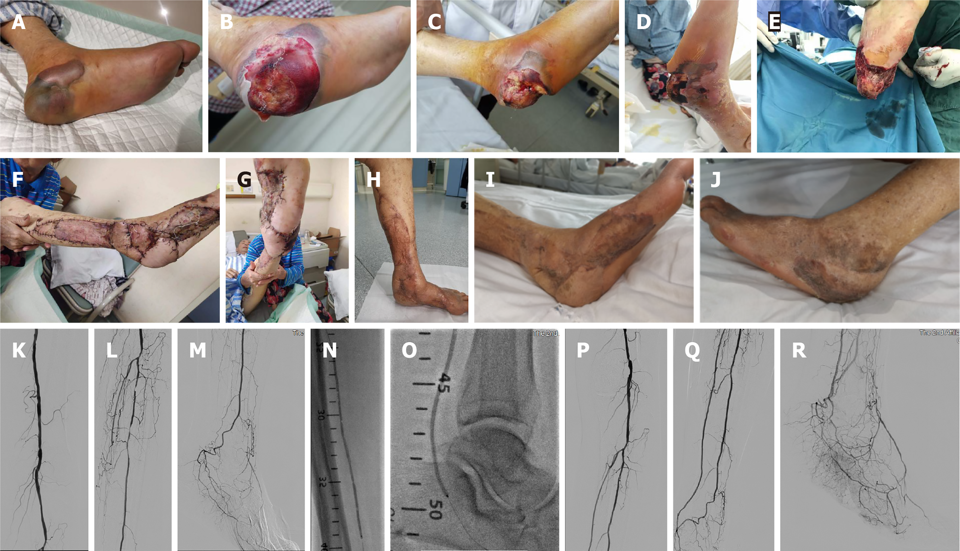
Figure 4 Surgery pictures of a local infection case.
A-D: The heel of a diabetic foot patient was punctured by a nail, resulting in local infection and gradual degeneration and necrosis of the soft tissue of the heel; E-G: Complete debridement was performed after revascularization, and the heel was repaired with a free-style perforator flap on the lower leg side; H-J: Two months after surgery, the patient's foot was successfully preserved and functioned well; K-M: Angiography showed patency of the femoral-popliteal artery, occlusions of the infrapopliteal and anterior tibial veins, occlusion of the middle and distal posterior tibial artery, and staged stenosis of peroneal artery; N and O: balloon angioplasty (BA) of posterior tibial artery and peroneal artery was performed; P-R: Angiography after BA showed that the blood flow of posterior tibial artery and peroneal artery was smooth with the blood flow directly to the heel, and the plantar arterial arch was good.
- Citation: Lei FR, Shen XF, Zhang C, Li XQ, Zhuang H, Sang HF. Clinical efficacy of endovascular revascularization combined with vacuum-assisted closure for the treatment of diabetic foot. World J Diabetes 2024; 15(7): 1499-1508
- URL: https://www.wjgnet.com/1948-9358/full/v15/i7/1499.htm
- DOI: https://dx.doi.org/10.4239/wjd.v15.i7.1499


