Copyright
©The Author(s) 2024.
World J Diabetes. Jun 15, 2024; 15(6): 1317-1339
Published online Jun 15, 2024. doi: 10.4239/wjd.v15.i6.1317
Published online Jun 15, 2024. doi: 10.4239/wjd.v15.i6.1317
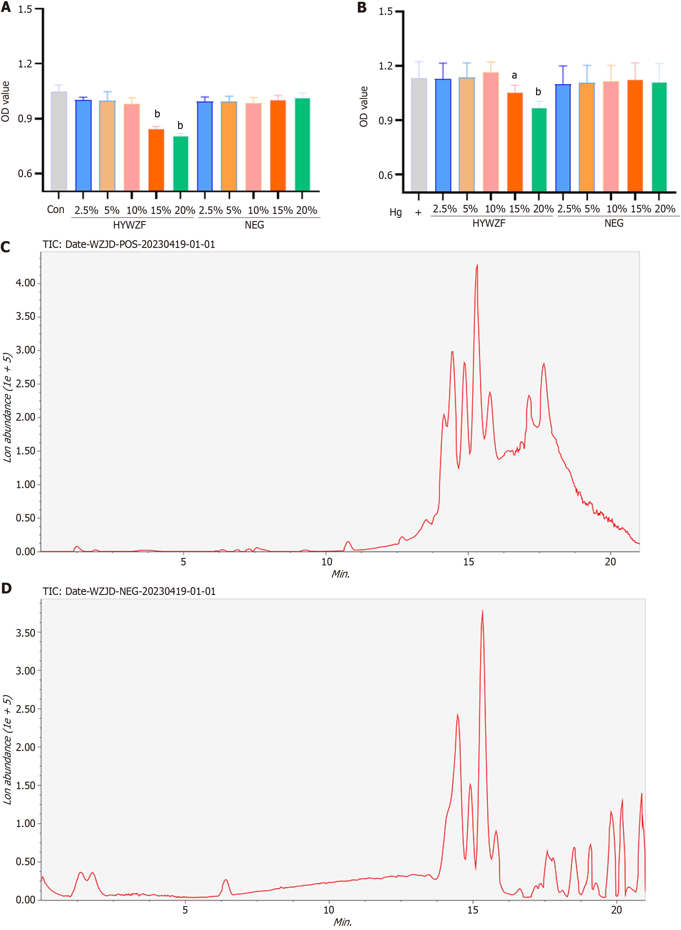
Figure 1 The analysis of heying wuzi formulation serum effects on human retinal capillary endothelial cell viability in high glucose Dulbecco’s modified Eagle’s medium and active ingredients.
A: CCK-8 assay was performed to determine the heying wuzi formulation (HYWZF) serum effects on cell viability in ordinary Dulbecco’s modified Eagle’s medium (DMEM, D-glucose, 5.5 mM); B: HYWZF serum effects on cell viability in high glucose DMEM (D-glucose, 33 mM); C: Chromatographic analysis for HYWZF (positive ion mode); D: Chromatographic analysis for HYWZF (negative ion mode). aP value < 0.05, bP value < 0.01. HYWZF: Heying wuzi formulation; NEG: Negative serum.
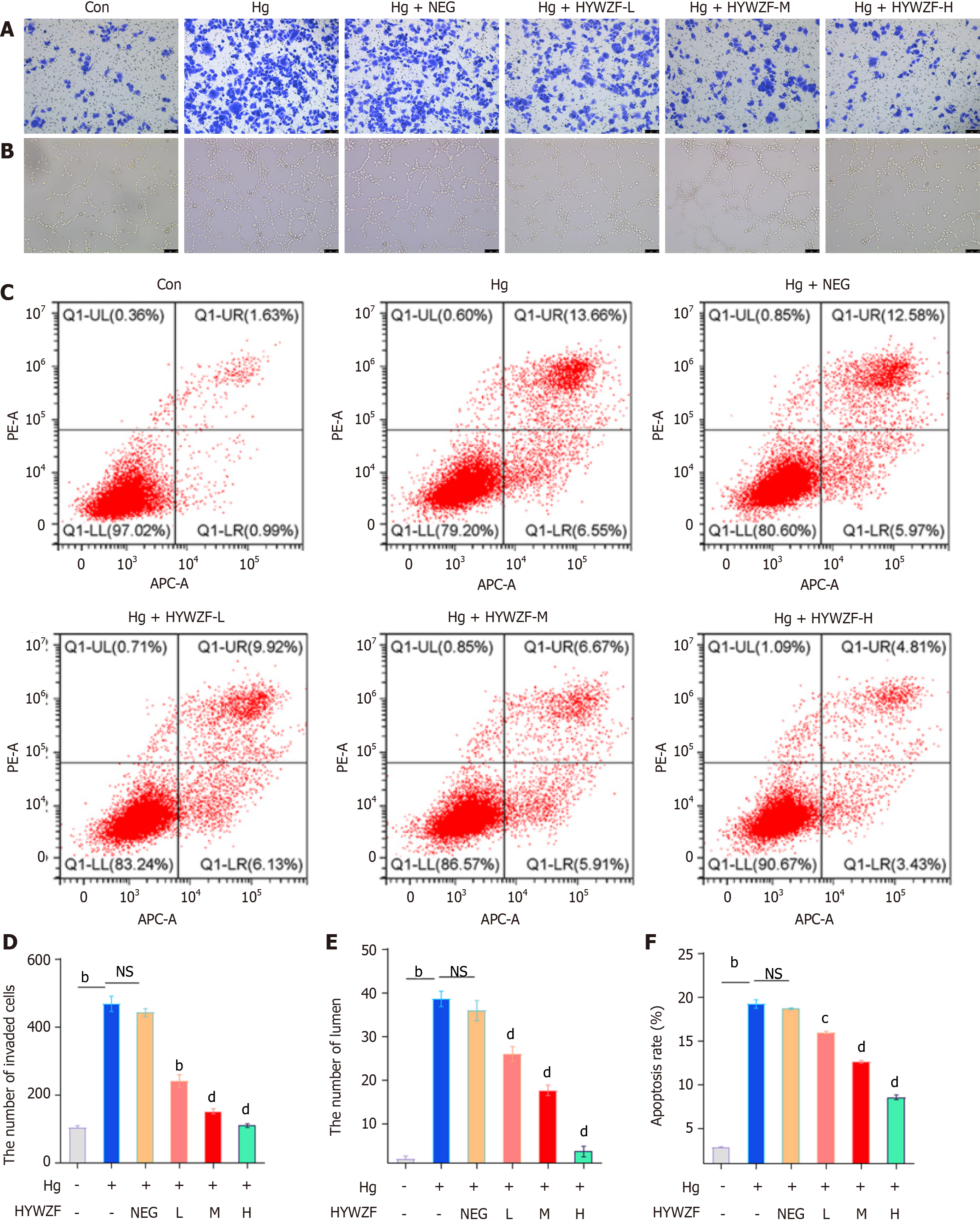
Figure 2 Impact of heying wuzi formulation serum on the invasion, phenotype, and apoptosis of human retinal capillary endothelial cells.
A: Transwell assay was used to detect cell invasion (scale bar: 100 μm); B: Tube formation assay detected the tube-forming ability (scale bar: 100 μm); C: Cell apoptosis was detected by flow cytometry; D: The number of invaded cells; E: The number of lumen formation of human retinal capillary endothelial cell (HRCEC); F: The apoptosis rate of HRCEC cells. Compared with the high glucose (hg) group, aP value < 0.05, bP value < 0.01; compared with the hg + negative serum group, cP value < 0.05, dP value < 0.01. HYWZF: Heying wuzi formulation; NEG: Negative serum; hg: High glucose; Con: Control.
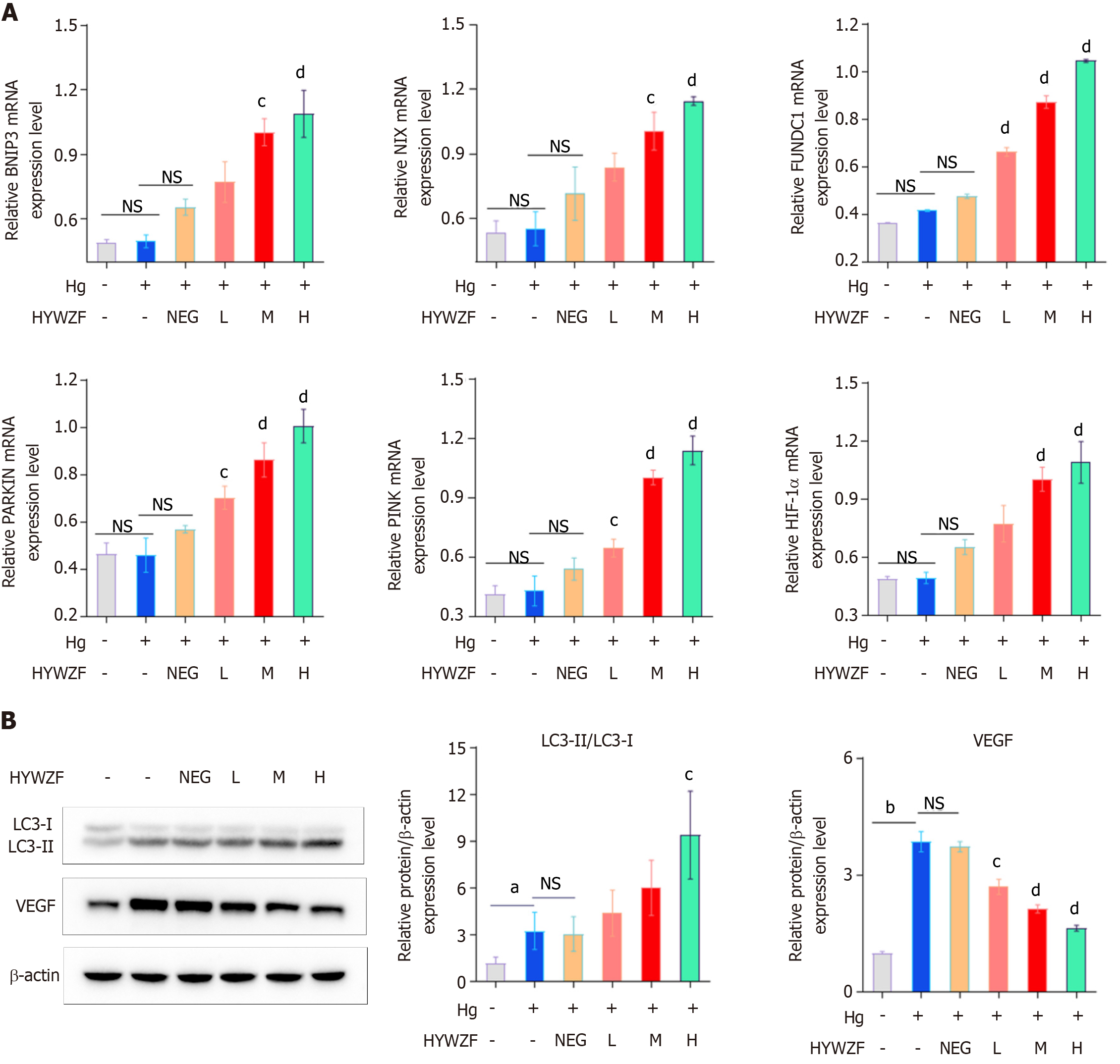
Figure 3 Mitophagy of human retinal capillary endothelial cells analysis.
A: Quantitative reverse transcription-polymerase chain reaction detection results of relative BNIP3, FUNDC1, NIX, PARKIN, PINK1, and HIF-1α mRNA expression levels; B: Western blotting analyzed the protein levels of light chain 3 (LC3)-I, LC3-II, and vascular endothelial cell growth factor. Compared with the high glucose (hg) group, aP value < 0.05, bP value < 0.01; compared with the hg + negative serum group, cP value < 0.05, dP value < 0.01. HYWZF: Heying wuzi formulation; NEG: Negative serum; hg: High glucose; VEGF: Vascular endothelial cell growth factor.
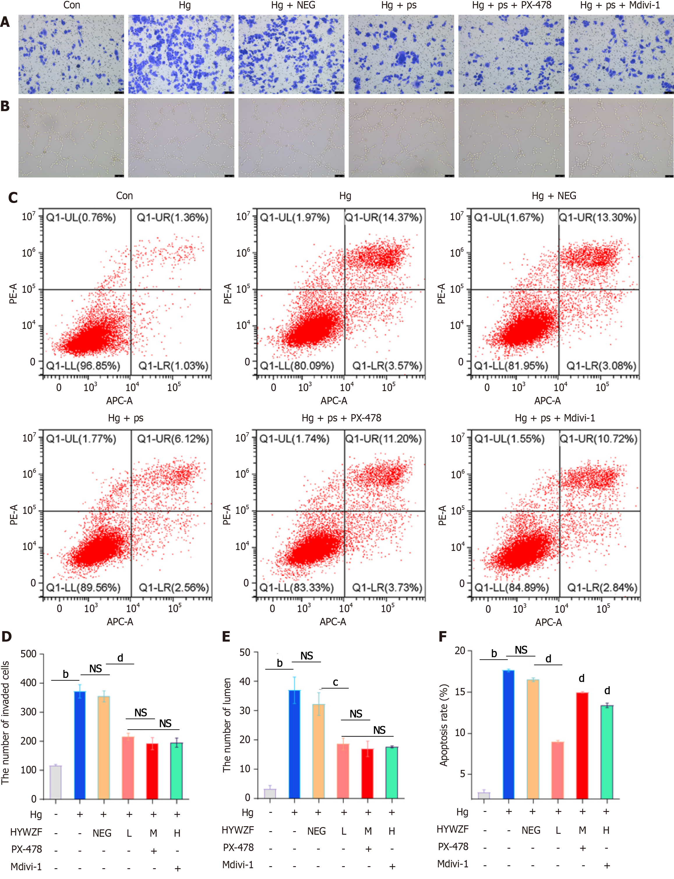
Figure 4 Heying wuzi formulation improved intracellular oxidative stress.
A and D: A transwell was performed to detect the invasion ability of Human retinal capillary endothelial cell (HRCEC; scar bar: 100 μm); B and E: A tube formation assay was performed to detect the tube-forming ability of HRCEC cells (scar bar: 100 μm); C and F: Cell apoptosis was detected by flow cytometry. Compared with the high glucose (hg) group, aP value < 0.05, bP value < 0.01; compared with the hg + 10% HYWZF serum group, cP value < 0.05, dP value < 0.01. HYWZF: Heying wuzi formulation; NEG: Negative serum; hg: High glucose; ps: 10% HYWZF serum; Con: Control.
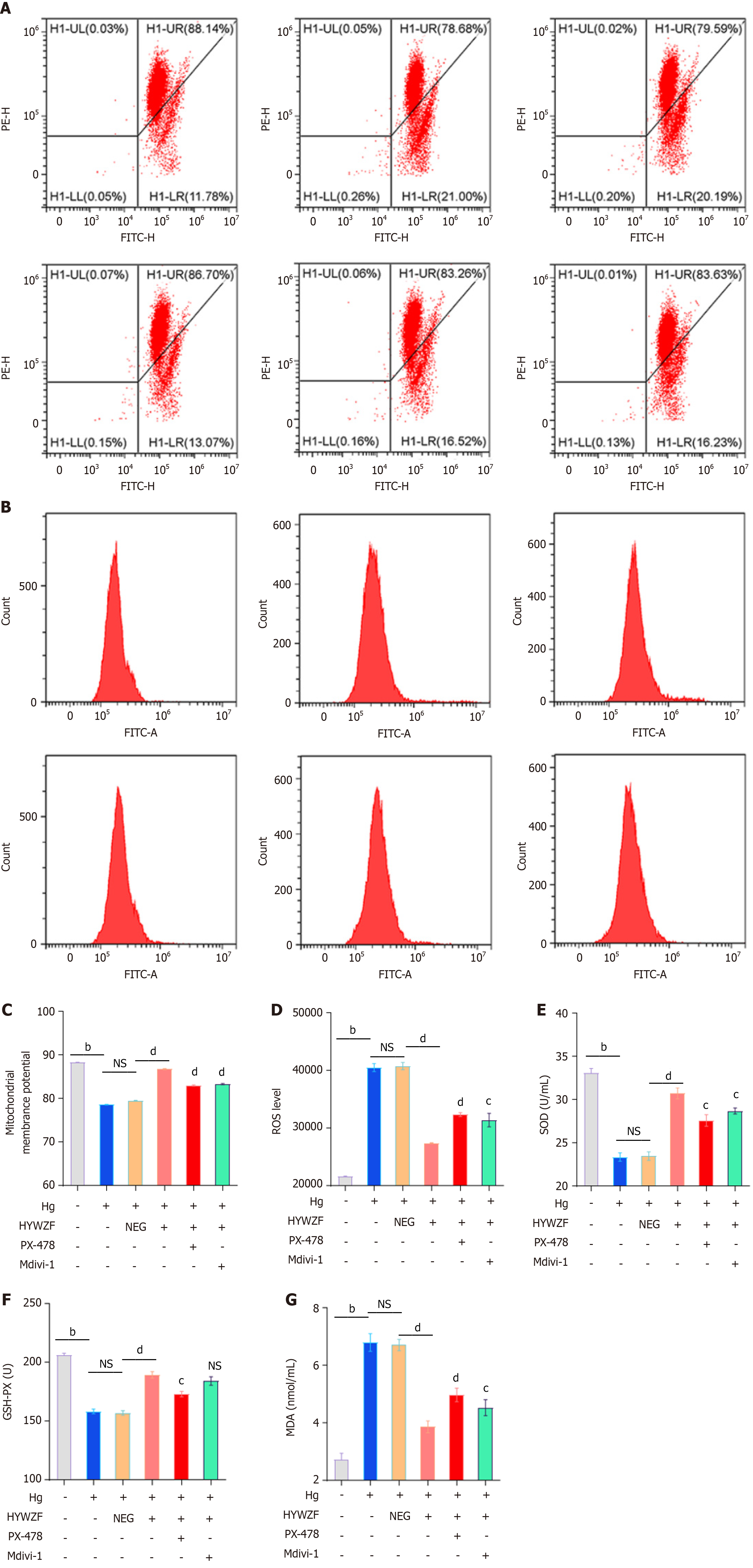
Figure 5 Effects of heying wuzi formulation on mitochondria and oxidative stress.
A and C: JC-1 detected mitochondrial membrane potential; B and D: reactive oxygen species was detected by flow cytometry; E-G: Enzymatic reaction detection of SOD, GSH-PX, and MDA levels. Compared with the high glucose (hg) group, aP value < 0.05, bP value < 0.01; compared with the hg + 10% heying wuzi formulation serum group, cP value < 0.05, dP value < 0.01. HYWZF: Heying wuzi formulation; mod: Diabetic model; NEG: Negative serum; hg: High glucose; ps: 10% HYWZF serum; Con: Control.
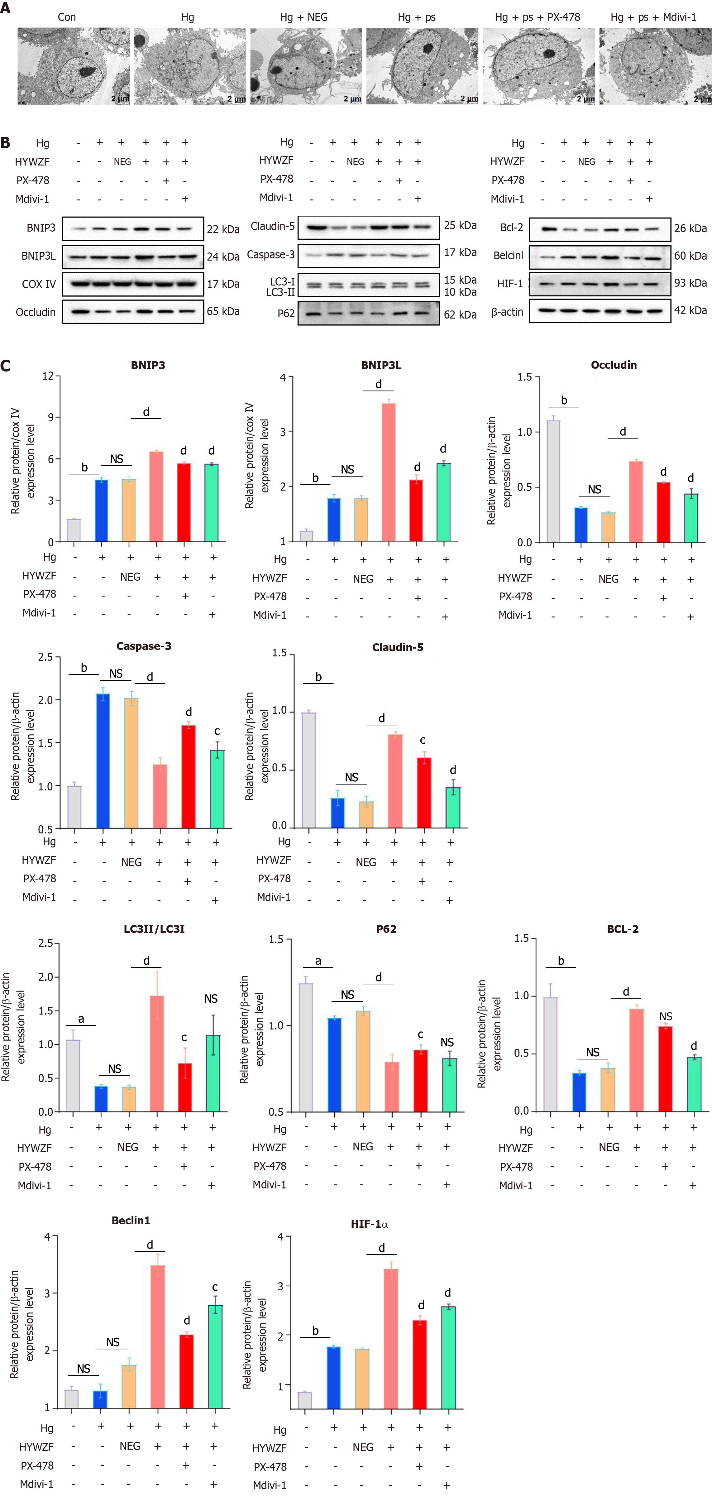
Figure 6 The role of heying wuzi formulation on mitochondrial morphology and function.
A: Morphology of mitochondria examined by TEM; B and C: Western blotting analysis of protein level. Compared with the high glucose (hg) group, aP value < 0.05, bP value < 0.01; compared with the hg + 10% Heying wuzi formulation serum group, cP value < 0.05, dP value < 0.01. HYWZF: Heying wuzi formulation; NEG: Negative serum; hg: High glucose; ps: 10% HYWZF serum; Con: Control.
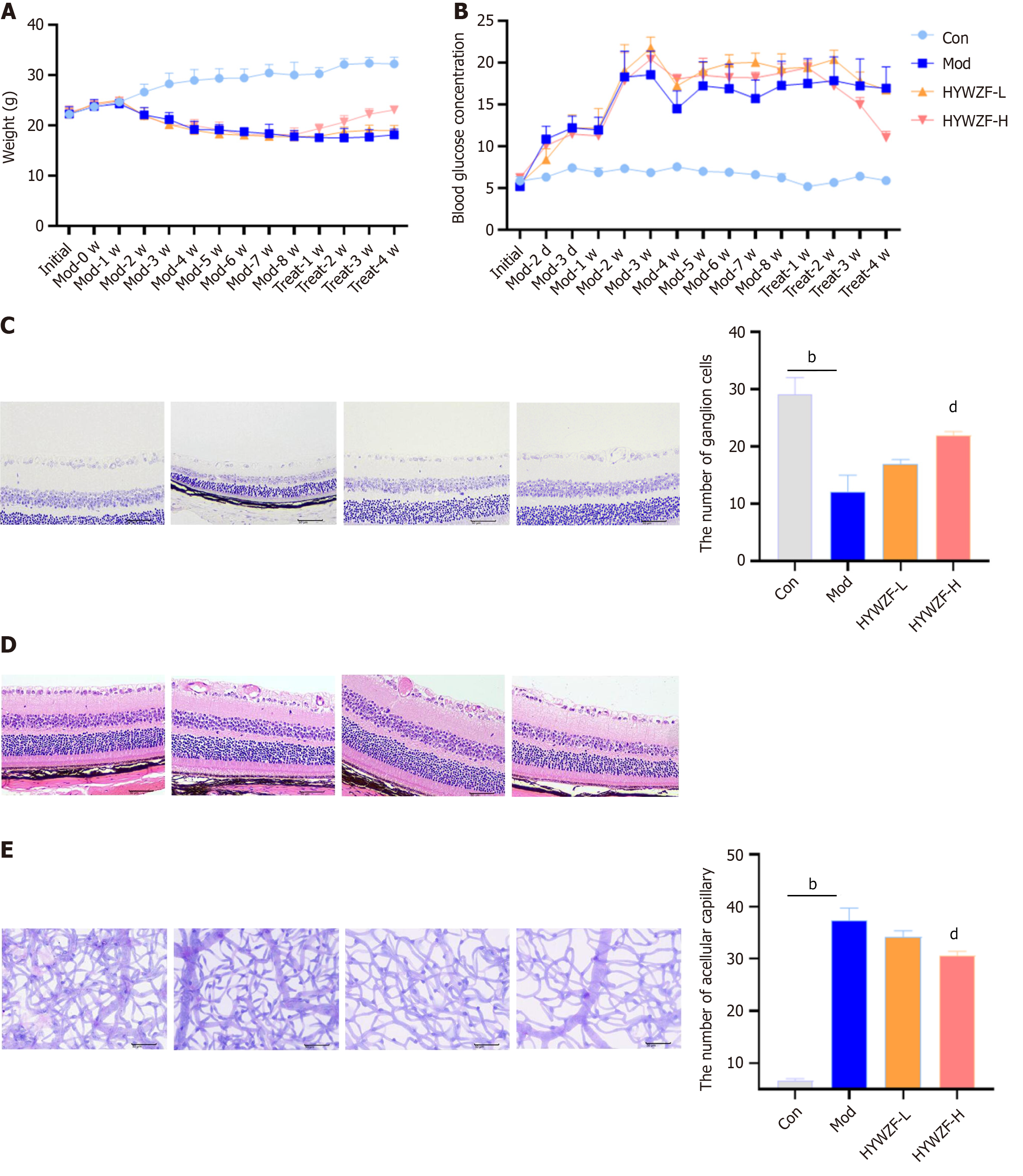
Figure 7 Heying wuzi formulation reduced pathological damage to retinal tissues.
A: The impact of heying wuzi formulation (HYWZF) on weight; B: The impact of HYWZF on blood glucose changes; C: The number of ganglion cells in retinal tissues by tar violet staining (scar bar: 50 μm); D: Hematoxylin and eosin staining was used to observe retinal tissue structure (scar bar: 50 μm); E: Periodic acid-shiff staining was used to detect the number of acellular capillaries in retinal tissues (scar bar: 50 μm). Compared with the con group, bP value < 0.01; compared with the diabetic model group, dP value < 0.01. HYWZF: Heying wuzi formulation; mod: Diabetic model; Con: Control.
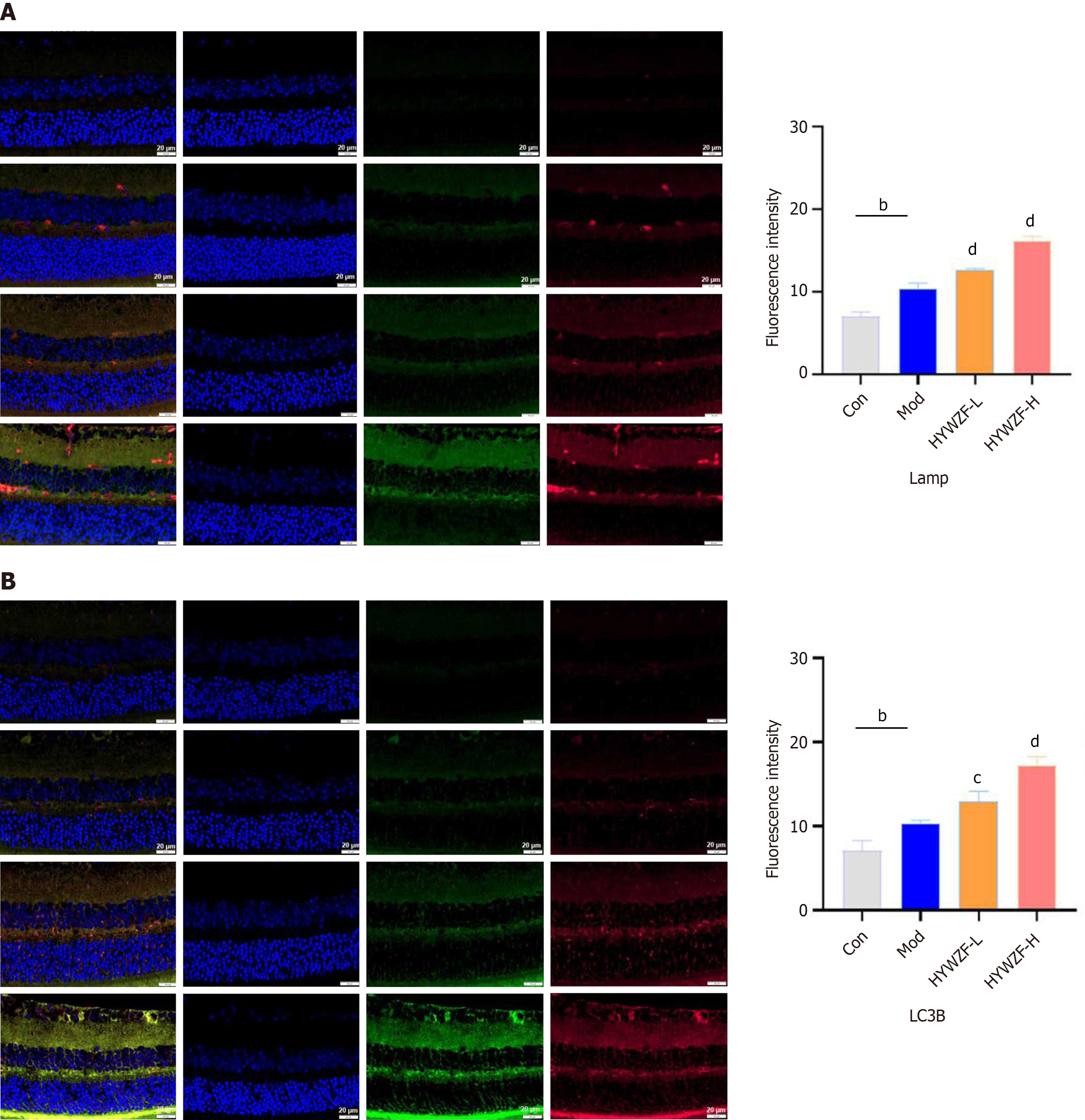
Figure 8 Heying wuzi formulation increased mitophagy.
A: Co-localization results of Tom20 and LAMP 2; B: Co-localization results of Tom20 and light chain 3B. Compared with the con group, bP value < 0.01; compared with the mod group, dP value < 0.01. HYWZF: Heying wuzi formulation; mod: Diabetic model; Con: Control.
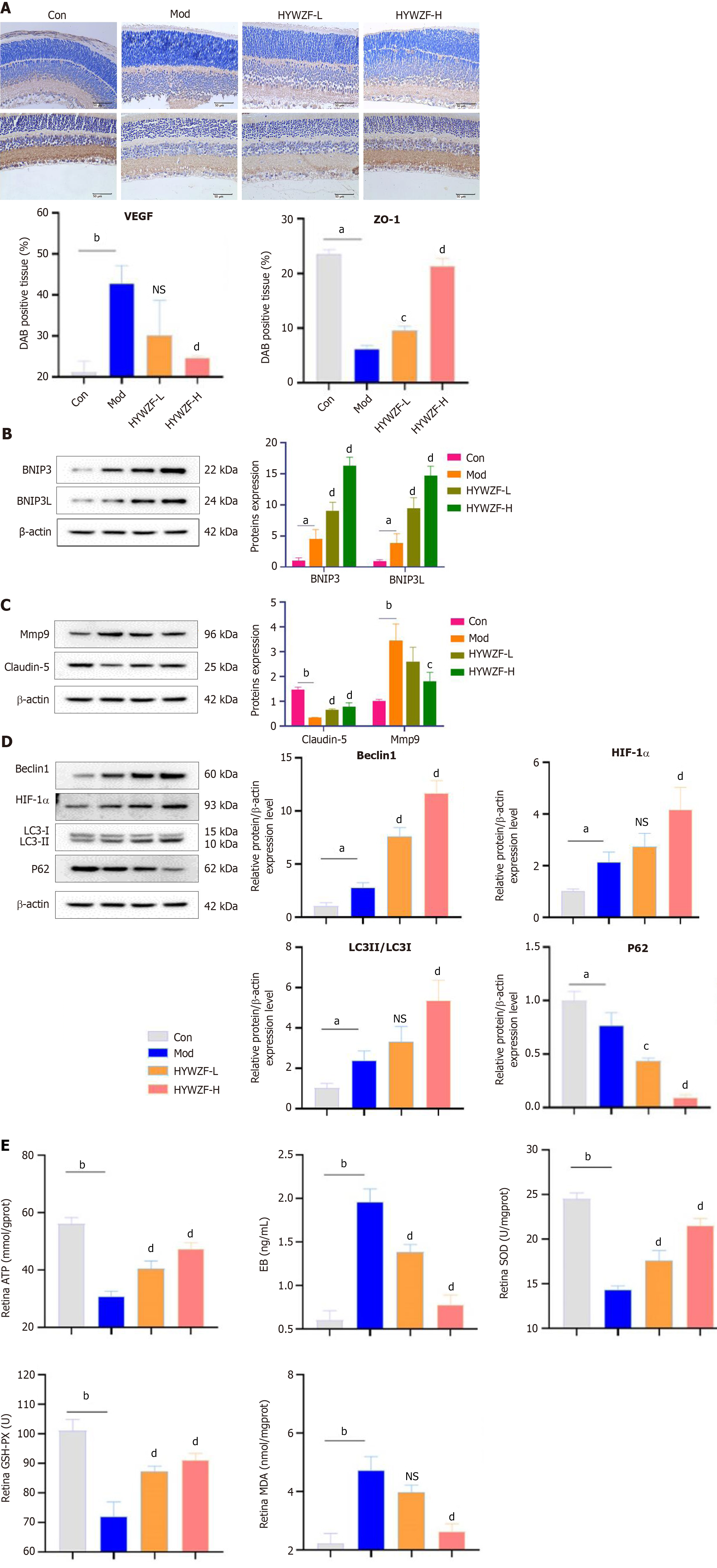
Figure 9 Effects of heying wuzi formulation on C57BL/6 mice.
A: Immunohistochemical was performed to quantify the protein expression of vascular endothelial cell growth factor and ZO-1; B-D: Western blotting analysis of protein levels of light chain 3B, BNIP3, HIF-1α, mmp9, and p62, etc. in retinal tissues; E: Enzymatic reaction detection of SOD, GSH-PX, ATP, EB, and MDA levels in the retina. Compared with the control group, aP value < 0.05, bP value < 0.01; compared with the diabetic model group, cP value < 0.05, dP value < 0.01. HYWZF: Heying wuzi formulation; VEGF: Vascular endothelial cell growth factor.
- Citation: Wu JJ, Zhang SY, Mu L, Dong ZG, Zhang YJ. Heyingwuzi formulation alleviates diabetic retinopathy by promoting mitophagy via the HIF-1α/BNIP3/NIX axis. World J Diabetes 2024; 15(6): 1317-1339
- URL: https://www.wjgnet.com/1948-9358/full/v15/i6/1317.htm
- DOI: https://dx.doi.org/10.4239/wjd.v15.i6.1317


