Copyright
©The Author(s) 2023.
World J Diabetes. Sep 15, 2023; 14(9): 1349-1368
Published online Sep 15, 2023. doi: 10.4239/wjd.v14.i9.1349
Published online Sep 15, 2023. doi: 10.4239/wjd.v14.i9.1349
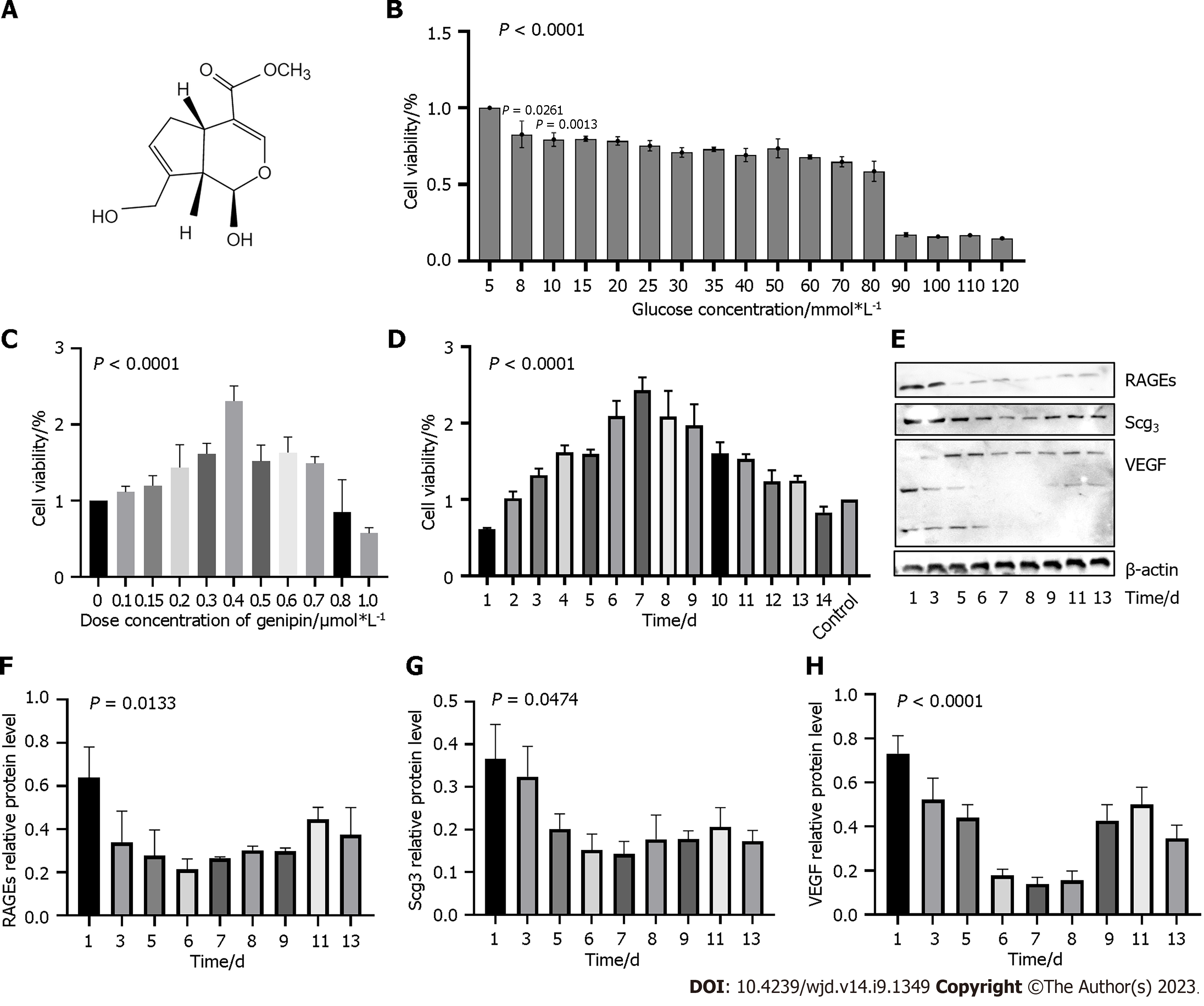
Figure 1 Optimization of dose of genipin and high glucose in human retinal microvascular endothelial cells.
A: Chemical structure of genipin; B and C: Cell viability of human retinal microvascular endothelial cells (hRMECs) treated with different concentrations of glucose and genipin; D: Cell viability of hRMECs treated with 30 mmol/L glucose and 0.4 μmol/L genipin for different durations; E-I: RAGE, SCG3, and vascular endothelial growth factor protein expression in cells treated with genipin for different durations. VEGF: Vascular endothelial growth factor.
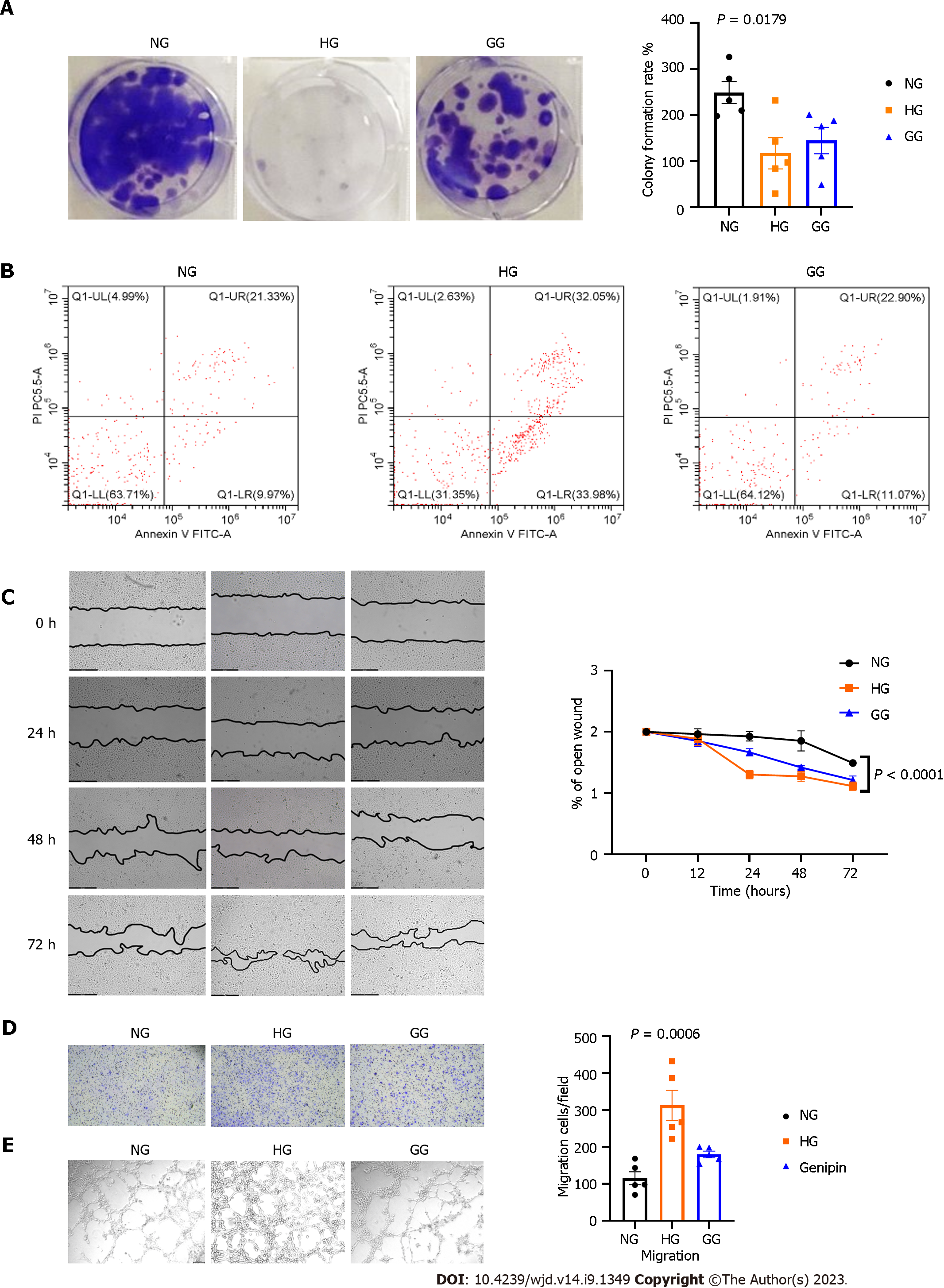
Figure 2 Genipin protects from advanced glycation-induced human retinal microvascular endothelial cell proliferation, apoptosis, and angiogenesis.
A and B: Genipin protects human retinal microvascular endothelial cell (hRMECs) from high glucose-induced damage with regard to cell proliferation as revealed by colony formation assay and flow cytometry; C-E: Wounding healing, migration and tube formation of hRMECs treated with 30 mmol/L glucose and 0.4 μmol/L genipin. NG: 5 mmol/L glucose containing ECM-treated hRMECs group; HG: 30 mmol/L glucose containing ECM-treated hRMECs group; GG: 0.4 μM genipin + 30 mmol/L glucose containing ECM-treated hRMECs.
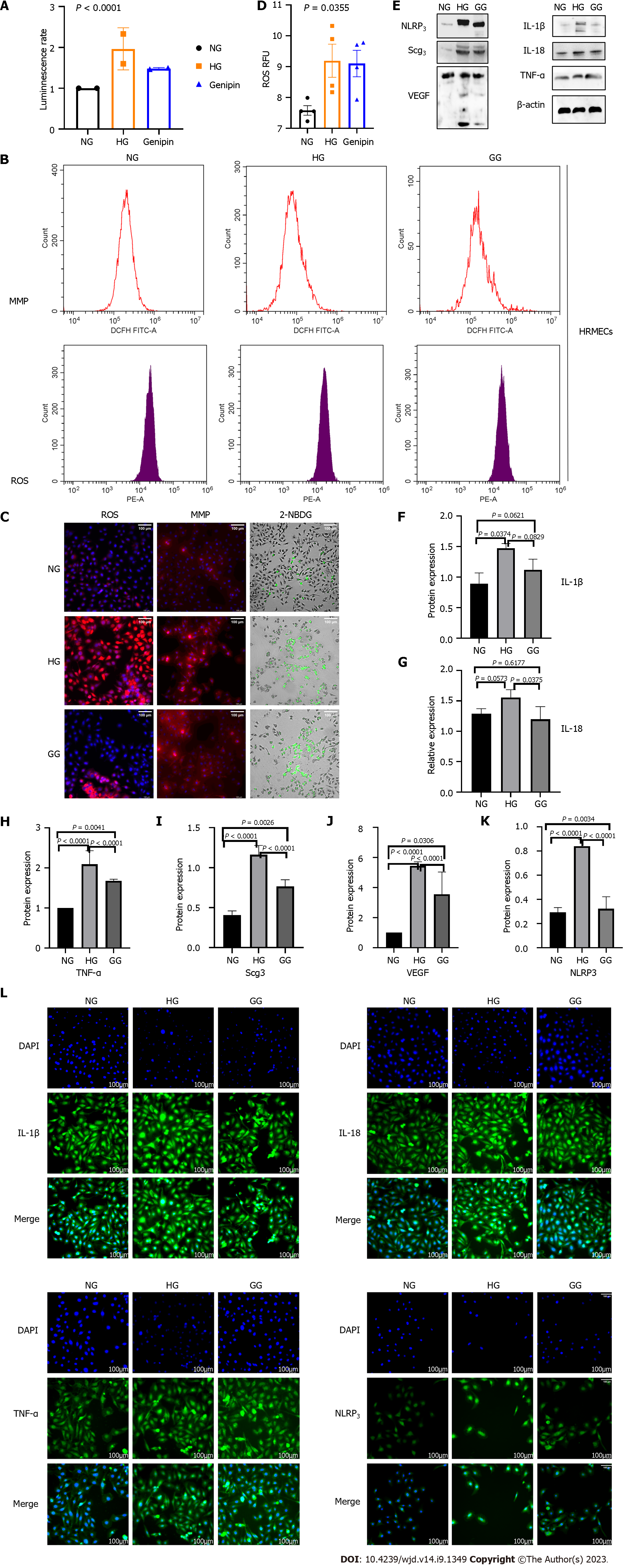
Figure 3 Genipin protects human retinal microvascular endothelial cells from high glucose damage with regard to energy metabolism, oxidative stress, and inflammatory injury in vitro.
A: ATP levels among normal glucose, high glucose, and genipin-treated human retinal microvascular endothelial cells (hRMECs); B-D: Reactive oxygen species, mitochondrial membrane potential, and 2-[N-(7-nitrobenz-2-oxa-1,3-diazol-4-yl) amino]-2-deoxy-d-glucose levels in hRMECs treated with high glucose and genipin; E-L: Western blot and immunofluorescence to measure the expression of inflammatory factors expression. IL: Interleukin; TNF: Tumor necrosis factor-alpha; NLRP3: Nucleotide-binding domain, leucine-rich-containing family, pyrin domain-containing 3; VEGF: Vascular endothelial growth factor; ROS: Reactive oxygen species; MMP: Mitochondrial membrane potential; 2-NBDG: 2-[N-(7-nitrobenz-2-oxa-1,3-diazol-4-yl) amino]-2-deoxy-d-glucose; hRMECs: Human retinal microvascular endothelial cells. NG: 5 mmol/L glucose containing ECM-treated hRMECs group; HG: 30 mmol/L glucose containing ECM-treated hRMECs group; GG: 0.4 μM genipin + 30 mmol/L glucose containing ECM-treated hRMECs.
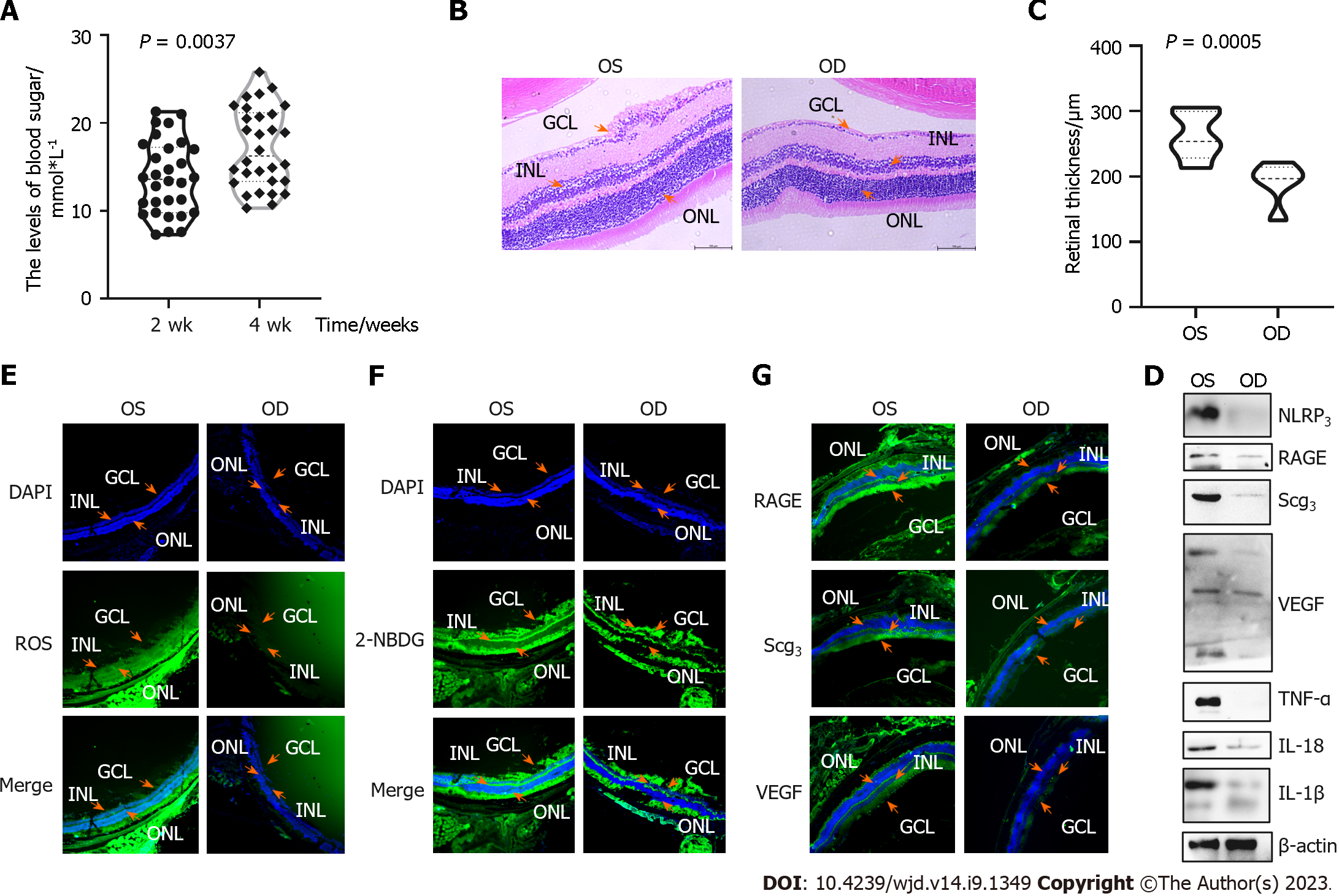
Figure 4 Genipin reduces retinal oxidant stress, glucose metabolism, and inflammation in streptozotocin-induced mice.
A: Blood sugar levels of mice induced with streptozotocin (STZ) for 2 wk and 4 wk; B: Hematoxylin and eosin staining of STZ mouse retina; C: Retinal thickness in different eyes of STZ mice; D: Inflammatory factor expression measured by Western blot; E and F: Reactive oxygen species and 2-[N-(7-nitrobenz-2-oxa-1,3-diazol-4-yl) amino]-2-deoxy-d-glucose levels in STZ mice; G and F: RAGE, SCG3, and vascular endothelial growth factor expression in STZ mice. IL: Interleukin; TNF: Tumor necrosis factor-alpha; NLRP3: Nucleotide-binding domain, leucine-rich-containing family, pyrin domain-containing 3; VEGF: Vascular endothelial growth factor; ROS: Reactive oxygen species; MMP: Mitochondrial membrane potential; 2-NBDG: 2-[N-(7-nitrobenz-2-oxa-1,3-diazol-4-yl) amino]-2-deoxy-d-glucose; OS: Left eye, without genipin-treated; OD: Right eye, genipin-treated; GCL: Ganglion cell layer; INL: Inner nuclear layer; ONL: Outer nuclear layer.
Figure 5 Mechanism of genipin to relive retinal endothelial damage caused by high glucose.
A and B: Schematic summary of the results; C-I: Western blot and immunofluorescence (IF) analysis of expression of proteins related to CHGA/UCP2/GLUT1; J: RAGE, SCG3, and vascular endothelial growth factor expression detected by IF.
Figure 6 Pathway enrichment analysis.
A: Prediction of diabetic retinopathy (DR)-related genes; B: Pathway enrichment analysis of DR-related genes; C and D: Intersection of genipin target and DR-related genes; E: Pathway enrichment analysis of genes which are mixed by DR-related and genipin targets. EPO: Erythropoietin; BP: Biological process-pathways and larger processes made up of the activities of multiple gene products; MF: Molecular Function-molecular activities of gene products; KEGG: Kyoto Encyclopedia of Genes and Genomes.
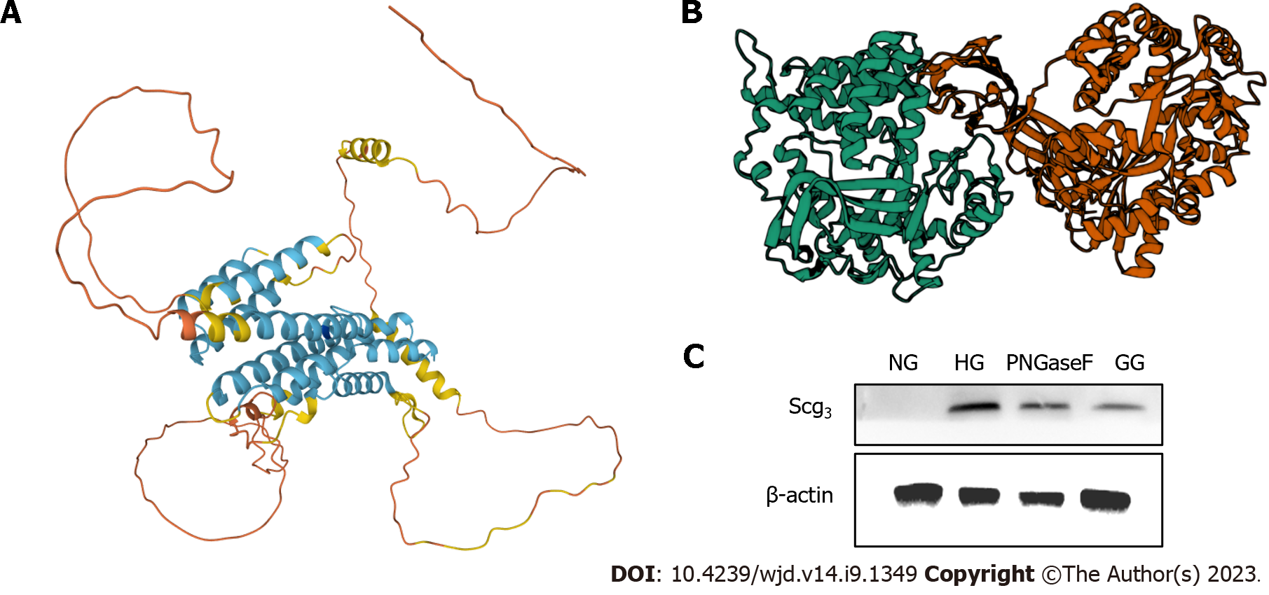
Figure 7 SCG3 plays a vital role in diabetic retinopathy via glycosylation.
A: Structure of SCG3; B: Molecular docking simulation with RAGE (green, left) and SCG3 (orange, right); C: PNGase F cleavaged Scg3. NG: 5 mmol/l glucose containing ECM-treated human retinal microvascular endothelial cells (hRMECs) group; HG: 30 mmol/L glucose containing ECM-treated hRMECs group; GG: 0.4 μM genipin + 30 mmol/L glucose containing ECM-treated hRMECs.
- Citation: Sun KX, Chen YY, Li Z, Zheng SJ, Wan WJ, Ji Y, Hu K. Genipin relieves diabetic retinopathy by down-regulation of advanced glycation end products via the mitochondrial metabolism related signaling pathway. World J Diabetes 2023; 14(9): 1349-1368
- URL: https://www.wjgnet.com/1948-9358/full/v14/i9/1349.htm
- DOI: https://dx.doi.org/10.4239/wjd.v14.i9.1349


