Copyright
©The Author(s) 2022.
World J Diabetes. Jun 15, 2022; 13(6): 434-441
Published online Jun 15, 2022. doi: 10.4239/wjd.v13.i6.434
Published online Jun 15, 2022. doi: 10.4239/wjd.v13.i6.434
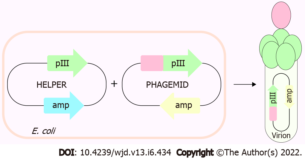
Figure 1 Depiction of phage display 3 + 3 system.
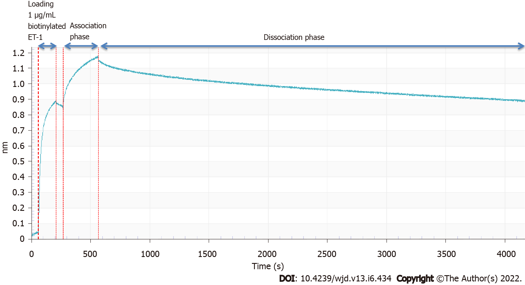
Figure 2 Sensorgram of Fc-β binding to biotinylated endothelin-1.
Representative plot shows the binding assay revealed that our selected endothelin (ET)-traps bind strongly to ET-1, displaying double-digit picomolar binding affinity (an average of 73.8 picoMolar, n = 6).
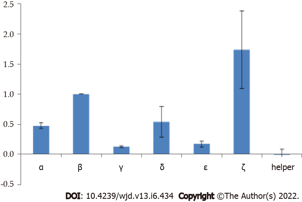
Figure 3 Average relative endothelin-1 binding of endothelin-traps constructs expressing clones using phage display.
The final signals are blank-subtracted and normalized to individual expression levels.
- Citation: Jain A, Bozovicar K, Mehrotra V, Bratkovic T, Johnson MH, Jha I. Investigating the specificity of endothelin-traps as a potential therapeutic tool for endothelin-1 related disorders. World J Diabetes 2022; 13(6): 434-441
- URL: https://www.wjgnet.com/1948-9358/full/v13/i6/434.htm
- DOI: https://dx.doi.org/10.4239/wjd.v13.i6.434


