Copyright
©The Author(s) 2021.
World J Diabetes. Dec 15, 2021; 12(12): 2011-2026
Published online Dec 15, 2021. doi: 10.4239/wjd.v12.i12.2011
Published online Dec 15, 2021. doi: 10.4239/wjd.v12.i12.2011
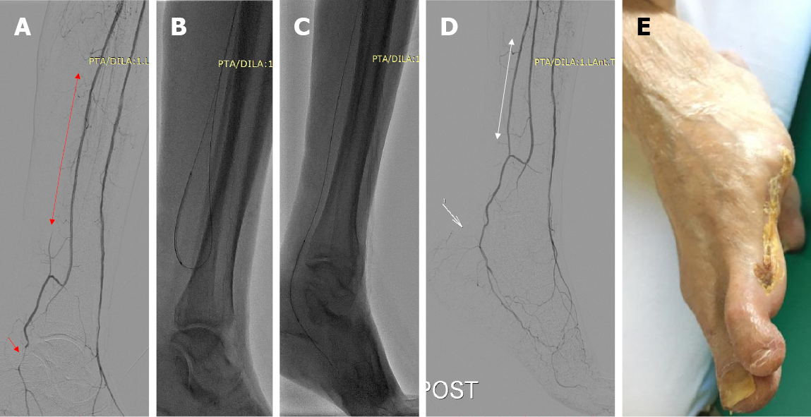
Figure 1 Wound-directed revascularization.
An 81-year-old female patient with long-standing type II diabetes and non-healing wound following minor amputation of the 3rd, 4th, and 5th toe and respective metatarsals. A: Digital subtraction angiography (DSA) demonstrating patent anterior tibial and peroneal arteries, occlusion of the posterior tibial artery from its origin (red line with arrowheads), and significant stenosis of the distal below the ankle posterior tibial artery (red arrow), which supplies the area of the surgical wound. Note that wound healing was not satisfactory even though the anterior tibial artery was patent to the distal foot; B and C: Retrograde revascularization of the posterior tibial artery via the peroneal artery and balloon angioplasty followed by (C) antegrade balloon angioplasty of the below the ankle stenosis via the revascularized posterior tibial artery; D: Final DSA depicting excellent angiographic patency of the treated vessels; E: Complete wound healing noted at 3 mo follow-up.
- Citation: Spiliopoulos S, Festas G, Paraskevopoulos I, Mariappan M, Brountzos E. Overcoming ischemia in the diabetic foot: Minimally invasive treatment options. World J Diabetes 2021; 12(12): 2011-2026
- URL: https://www.wjgnet.com/1948-9358/full/v12/i12/2011.htm
- DOI: https://dx.doi.org/10.4239/wjd.v12.i12.2011


