Copyright
©The Author(s) 2020.
World J Diabetes. Dec 15, 2020; 11(12): 622-643
Published online Dec 15, 2020. doi: 10.4239/wjd.v11.i12.622
Published online Dec 15, 2020. doi: 10.4239/wjd.v11.i12.622
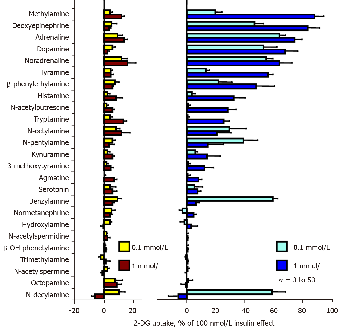
Figure 1 Screening of amines enhancing glucose transport in rat adipocytes under the influence of vanadate.
Suspensions of rat fat cells were incubated without (left) or with 100 µmol/L sodium orthovanadate (right) for 45 min in the presence of the indicated agents at 0.1 mmol/L (clearer columns) or 1 mmol/L (dark columns) just prior to the [3H]-2-deoxyglucose (2-DG) transport assay for 10 min. 2-DG uptake was expressed as the percentage of maximal stimulation by 100 nmol/L insulin, with baseline set at 0%. Rank order of the amines was based according to their effect at 1 mmol/L + 0.1 mmol/L vanadate. The mean ± SE from at least 3 determinations (for the agents almost inactive, at the bottom of the graph) up to 53 observations, for positive hits such as deoxyepinephrine, adrenaline (epinephrine), noradrenaline (norepinephrine), dopamine, methylamine, tyramine and benzylamine, with the seven most efficient amines being significant at P < 0.001. 2-DG: 2-Deoxyglucose.
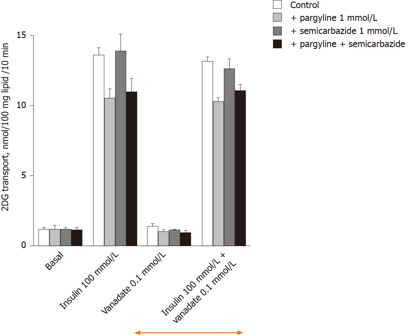
Figure 2 Lack of influence of amine oxidase inhibitors and of 100 µmol/L vanadate on basal and insulin-stimulated hexose uptake in rat adipocytes.
Rat fat cells (approximately 18 mg lipid/400 µL) were incubated for 45 min with the indicated concentrations of agents without (control, open columns), or with 1 mmol/L pargyline and 1 mmol/L semicarbazide, respectively (grey columns) or in combination (dark columns); then [3H]-2-deoxyglucose uptake (2-DG) was assayed over a 10-min period. The mean ± SE of 4 separate experiments was determined. There was no significant difference between inhibitors and each respective control. Similarly, no difference was found between corresponding conditions (basal or 100 nmol/L insulin-stimulated 2-DG transport) without (left) and with 100 µmol/L vanadate (right, above orange arrow) by the paired t test. 2-DG: 2-Deoxyglucose.
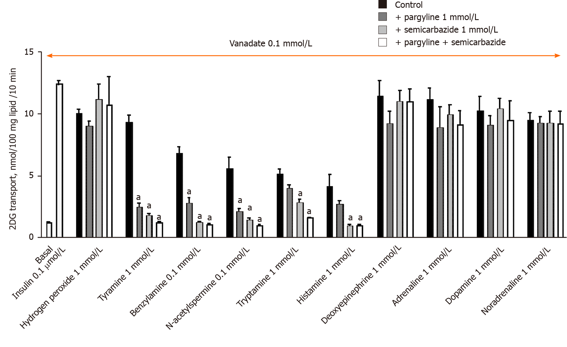
Figure 3 Inhibition by pargyline, semicarbazide, and their combination on amine-induced hexose uptake in rat adipocytes in the presence of 100 µmol/L sodium orthovanadate.
Immediately prior to the 10-min 2-deoxyglucose (2-DG) uptake assay, rat fat cells were incubated for 45 min as in Figure 2 in the presence of 100 µmol/L vanadate (orange arrow) without (basal) and with 100 nmol/L insulin (light grey columns), or with the indicated agents (control, black columns). 1 mmol/L pargyline or 1 mmol/L semicarbazide (grey columns) and their combination (open columns) were added to each agent (hydrogen peroxide or amine) tested at 1 mmol/L, except benzylamine, which was tested at 0.1 mmol/L. A significant reduction of 2-DG uptake when compared to the respective control was observed at: aP < 0.02. The mean ± SE of 4 adipocyte preparations was determined. 2-DG: 2-Deoxyglucose.
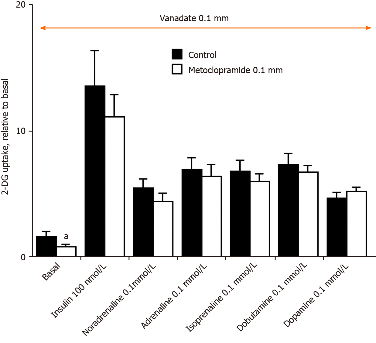
Figure 4 Lack of influence of the dopaminergic antagonist metoclopramide on insulin- or amine-stimulated hexose uptake in rat adipocytes.
Rat fat cells were incubated for 45 min with the indicated concentrations of agents without (control, closed columns), or with 0.1 mmol/L metoclopramide (open columns); then uptake of the glucose analogue 2-deoxyglucose (2-DG) was assayed over 10 min. 2-DG uptake was expressed as the fold increase relative to basal uptake set at 1.0. The mean ± SE of 4 separate experiments performed in the presence of 100 µmol/L vanadate (orange arrow) was determined. A significant difference between metoclopramide and the respective control was observed at: aP < 0.05. 2-DG: 2-Deoxyglucose.
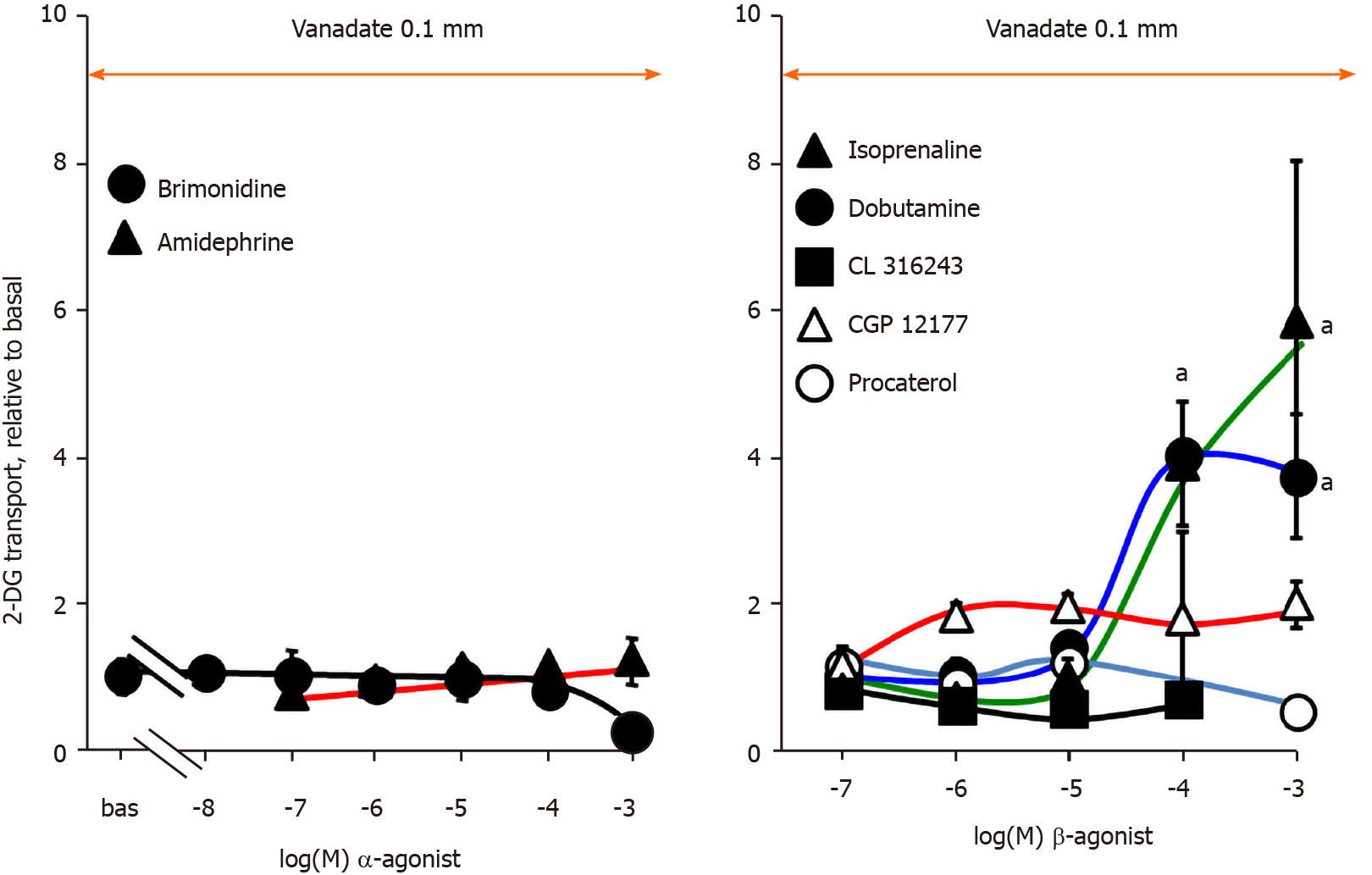
Figure 5 Effects of α- and β-adrenergic agonists on glucose transport in rat adipocytes.
2-Deoxyglucose uptake assay was performed as in Figure 3 with 100 µmol/L vanadate (orange arrows) and increasing doses of the indicated α- (left panel) or β-adrenergic agonists (right panel). Glucose transport was expressed as the fold increase relative to basal uptake (bas) set at 1.0. The mean ± SE of 4-6 adipocyte preparations was determined. Only high doses of isoprenaline and dobutamine induced responses were significantly different from baseline at: aP < 0.05. 2-DG: 2-Deoxyglucose.
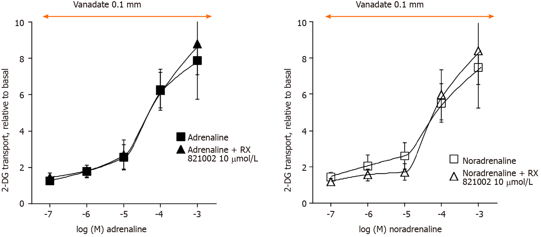
Figure 6 Dose-dependent activation by adrenaline and noradrenaline of hexose uptake in rat adipocytes: influence of the α2-adrenoreceptors antagonist methoxy-idazoxan.
Rat fat cells were incubated for 45 min with the indicated concentrations of adrenaline (left panel) or noradrenaline (right panel) without (control, squares), or with 0.01 mmol/L RX 821002 (triangles) prior to the 2-deoxyglucose uptake assay. Hexose transport was expressed as the fold increase relative to basal set at 1.0. The mean ± SE of 4 separate experiments performed in the presence of 100 µmol/L vanadate (orange arrow) was determined. No significant difference was found between the inhibitor and the respective control. 2-DG: 2-Deoxyglucose.
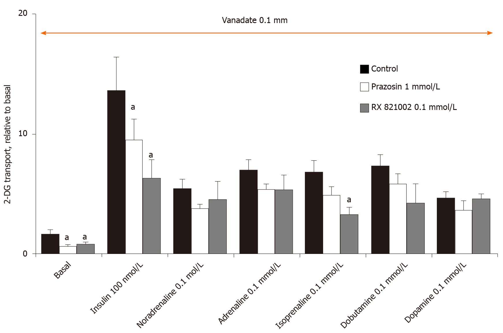
Figure 7 Inhibition of basal and insulin-stimulated hexose uptake in rat adipocytes by high doses of the α1-adrenoreceptors antagonist prazosin and the α2-adrenoreceptors antagonist methoxy-idazoxan.
Rat fat cells were incubated for 45 min with the indicated concentrations of agents without (control, closed columns), and with 1 mmol/L prazosin (open columns) or 0.1 mmol/L RX 821002 (shaded columns) prior to 2-deoxyglucose uptake, expressed as the fold increase relative to basal. The mean ± SE of 4 separate experiments performed in the presence of 100 µmol/L vanadate was determined. Significant difference between the inhibitor and the respective control was observed at: aP < 0.05. 2-DG: 2-Deoxyglucose.
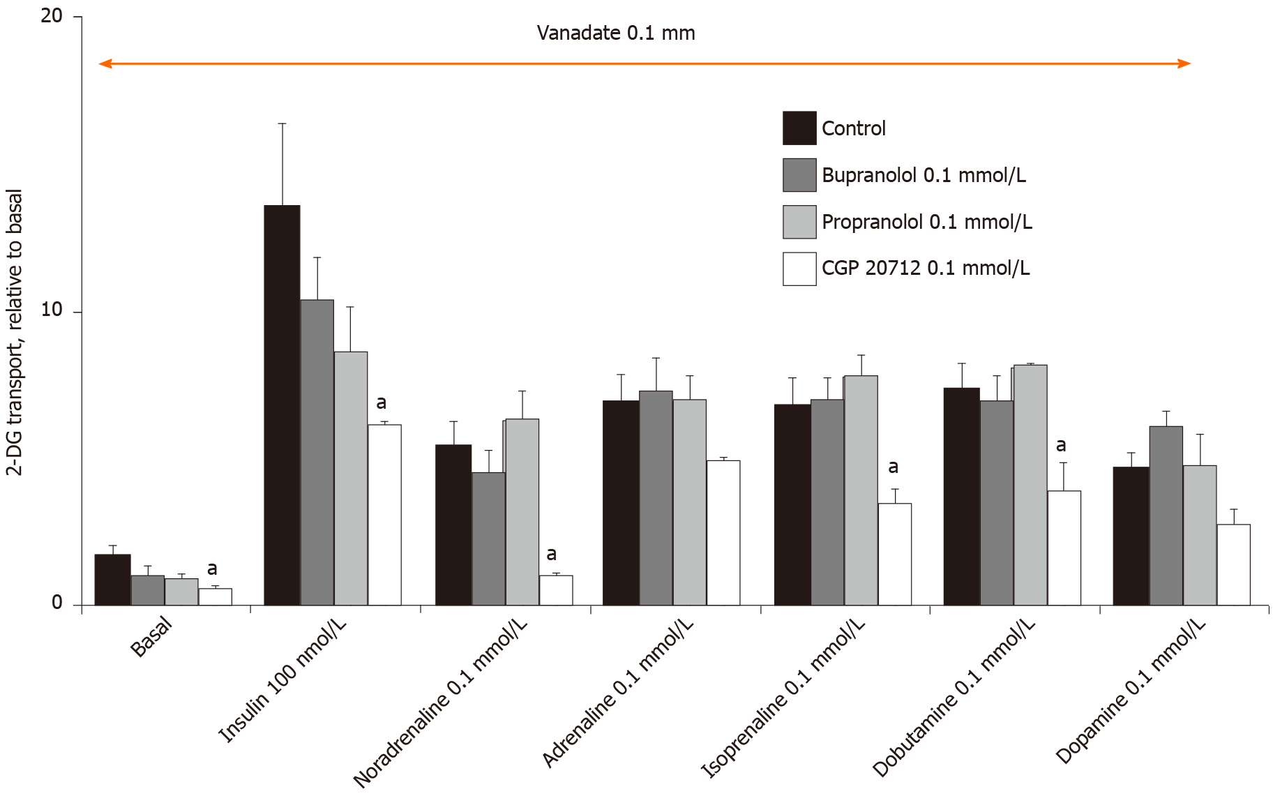
Figure 8 Influence of β-adrenoreceptors antagonists on hexose uptake in rat adipocytes.
Rat fat cells were incubated for 45 min with the indicated concentrations of agents without (control, closed columns), and with 0.1 mmol/L bupranolol (shaded columns), 0.1 mmol/L propranolol (grey columns) or 0.1 mmol/L CGP 20712 (open columns) prior to 2-deoxyglucose uptake, expressed as the fold increase relative to basal. The mean ± SE of 4 separate experiments performed in the presence of 100 µmol/L vanadate was determined. Significant difference between the inhibitor and the respective control was observed at: aP < 0.05. 2-DG: 2-Deoxyglucose.
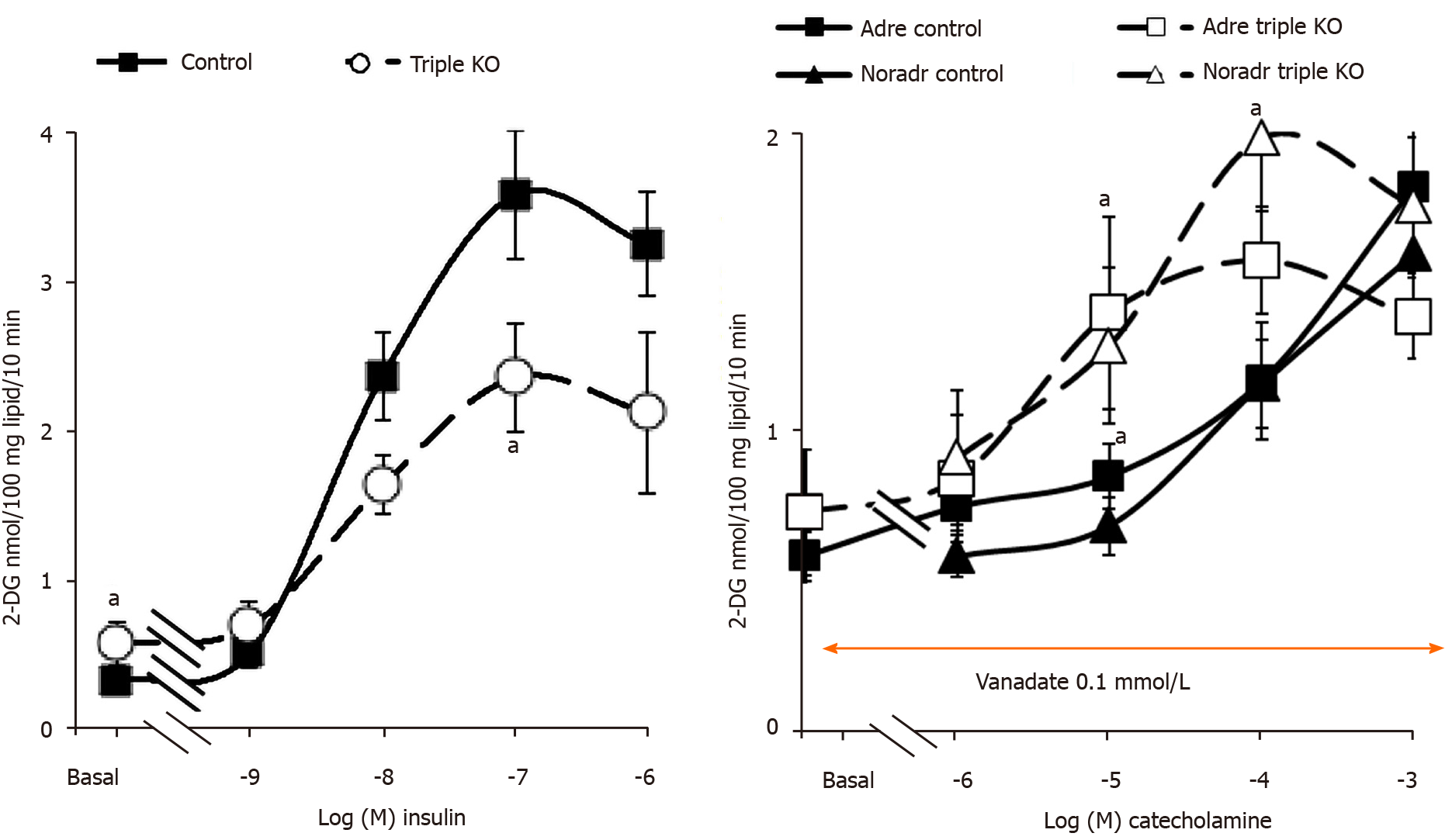
Figure 9 Glucose transport activation by insulin, adrenaline and noradrenaline in adipocytes from mice expressing or lacking β-adrenoceptors.
Left panel: Insulin dose-dependent stimulation of 2-deoxyglucose uptake in adipocytes from wild-type (control, black symbols) and "β-less" mice lacking β1-, β2-, β3-adrenoceptors (triple KO, open symbols). Right panel: Stimulation of hexose uptake by adrenaline (adre, squares) and noradrenaline (noradr, triangles) in the presence of 100 µmol/L vanadate (orange arrow). The mean ± SE of 4 (triple KO, dotted lines) to 13 (control, solid lines) adipocyte preparations was determined. A significant difference from the corresponding control was observed at: aP < 0.05. 2-DG: 2-Deoxyglucose.
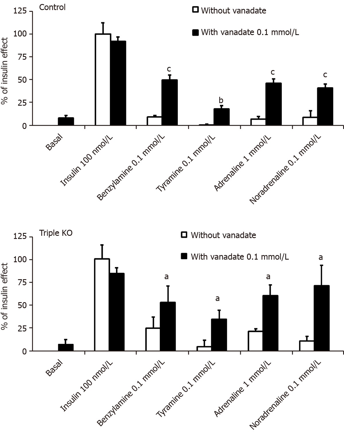
Figure 10 Influence of vanadate on hexose uptake into adipocytes from control and β-less mice.
Suspensions of mouse fat cells from control (upper panel) or mice with triple KO for the β1-, β2-, β3-adrenoceptors (lower panel) were incubated for 45 min without (basal, set at 0%), with 100 nmol/L insulin (set at 100%), or with the indicated doses of amines alone (open columns), or in combination with 100 µmol/L vanadate (dark columns), then [3H]-2-deoxyglucose uptake was assayed over 10 min. The mean ± SE of 4 (triple KO, lower panel) to 13 (control, upper panel) adipocyte preparations containing 17.2 ± 1.7 and 12.0 ± 1.6 mg lipids/400 µL, respectively, was determined. A significant difference from the corresponding condition without vanadate was observed at: aP < 0.05, bP < 0.01, cP < 0.001.
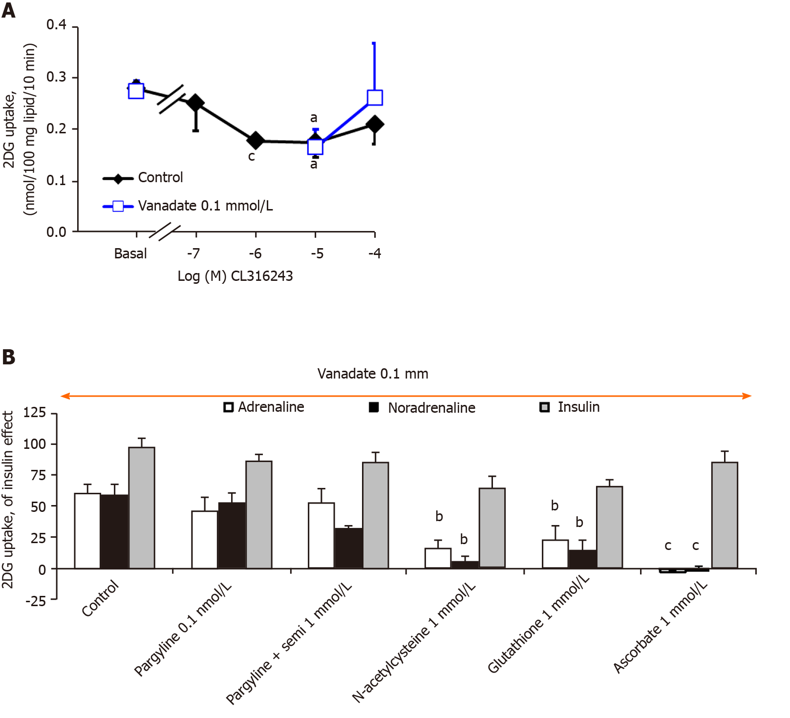
Figure 11 Influence of CL 316243, vanadate, amine oxidase inhibitors and antioxidants on glucose uptake in mouse adipocytes.
Fat cells from wild-type mice were subjected to 2-deoxyglucose (2-DG) uptake in the presence of CL 316243, or catecholamines, insulin and vanadate. A: The β3-adrenoceptors agonist was incubated alone (control, closed diamonds) or with vanadate (open squares) at the indicated concentrations; B: 1 mmol/L adrenaline (open columns) and noradrenaline (black columns) or 100 nmol/L insulin (shaded columns) were incubated with 100 µmol/L vanadate, without (control) or in the presence of pargyline 0.1 mmol/L, or pargyline + semicarbazide 1 mmol/L, or with 1 mmol/L of the antioxidants: N-acetylcysteine, glutathione, ascorbic acid. 2-DG uptake was expressed as the percentage of the insulin effect (which reached 1.73 ± 0.33 nmol 2-DG/100 mg lipids/0 min). The mean ± SE of 4 determination was determined. A significant difference from the respective control was observed at: aP < 0.05, bP < 0.01, cP < 0.001. 2-DG: 2-Deoxyglucose.
- Citation: Fontaine J, Tavernier G, Morin N, Carpéné C. Vanadium-dependent activation of glucose transport in adipocytes by catecholamines is not mediated via adrenoceptor stimulation or monoamine oxidase activity. World J Diabetes 2020; 11(12): 622-643
- URL: https://www.wjgnet.com/1948-9358/full/v11/i12/622.htm
- DOI: https://dx.doi.org/10.4239/wjd.v11.i12.622


