Copyright
©The Author(s) 2015.
World J Gastrointest Endosc. Nov 25, 2015; 7(17): 1257-1261
Published online Nov 25, 2015. doi: 10.4253/wjge.v7.i17.1257
Published online Nov 25, 2015. doi: 10.4253/wjge.v7.i17.1257
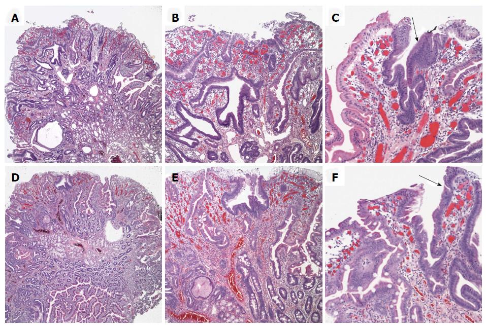
Figure 2 Histopathologic findings.
Biopsies from the mid duodenal bulb polyp showed villiform hyperplasia of intestinal and gastric foveolar epithelium with numerous capillaries demonstrating congestion and vascular ectasia (A and B). Similar changes seen in the polyp from the second part of the duodenum (D-E). The epithelium lining the surface and crypts focally (arrows) showing cells with mucin depletion and slightly pencillate nuclei with hyperchromasia (C and F). Representative images of hematoxylin and eosin stained slides taken at 40 × (A, D: 4 × objective and 10 × ocular magnification), 100 × (B, E: 10 × objective and 10 × ocular magnification) and 200 × (C, F: 20 × objective and 10 × ocular magnification).
- Citation: Gurung A, Jaffe PE, Zhang X. Duodenal polyposis secondary to portal hypertensive duodenopathy. World J Gastrointest Endosc 2015; 7(17): 1257-1261
- URL: https://www.wjgnet.com/1948-5190/full/v7/i17/1257.htm
- DOI: https://dx.doi.org/10.4253/wjge.v7.i17.1257


