Published online Mar 16, 2014. doi: 10.4253/wjge.v6.i3.99
Revised: November 9, 2013
Accepted: March 3, 2014
Published online: March 16, 2014
Processing time: 138 Days and 16.8 Hours
Neoplastic proliferation of plasma cells is called plasma cell dyscrasias, and these neoplasms can present as a solitary neoplasm or multiple myeloma. Extramedullary plasmacytoma, in particular pancreatic plasmacytoma, is a rare manifestation of multiple myeloma. Although computerized tomography is useful for the diagnosis of extramedullary plasmacytoma, there are no specific radiologic markers that distinguish it from adenocarcinoma. Histological confirmation by biopsy is necessary for accurate diagnosis and management of the tumor. Endosonography is the most sensitive method for the diagnosis of pancreatic tumors, and the use of fine needle aspiration by endosonography is associated with a lower risk for malignant seeding and complications. Here, we report a case of pancreatic plasmacytoma in newly identified multiple myeloma as diagnosed by endosonography. Endosonography is a reliable and rapid method for the diagnosis of extramedullary plasmacytoma. Therefore, endosonographic fine needle aspiration should be the first choice for histological evaluation when pancreatic plasmacytoma is suspected. Ideally, the pathology would be performed at the same site as endosonographic biopsy.
Core tip: The rare condition extramedullary plasmacytoma involves the gastrointestinal tract, usually liver, in approximately 10% of cases. A role for the pancreas is particularly rare. Pancreatic tumors can be identified radiologically, although it is impossible to discriminate between extramedullary plasmacytoma and adenocarcinoma. The use of endosonographic fine needle aspiration to acquire a histological sample from the pancreatic mass to confirm diagnosis is feasible and informative even in the presence of inoperable mass image.
- Citation: Akyuz F, Şahin D, Akyuz U, Vatansever S. Rare pancreas tumor mimicking adenocarcinoma: Extramedullary plasmacytoma. World J Gastrointest Endosc 2014; 6(3): 99-100
- URL: https://www.wjgnet.com/1948-5190/full/v6/i3/99.htm
- DOI: https://dx.doi.org/10.4253/wjge.v6.i3.99
An uncommon manifestation of multiple myeloma is extramedullary plasmacytoma. It is generally localized to nasal fossa and rarely involves the pancreas. On imaging, such a pancreatic mass may mimic adenocarcinoma[1,2].
We present here a case of a 64-year-old man who was referred to our endoscopy unit for endosonographic (EUS) fine needle aspiration (FNA) for a pancreatic mass.
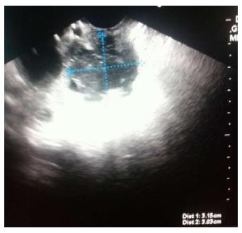
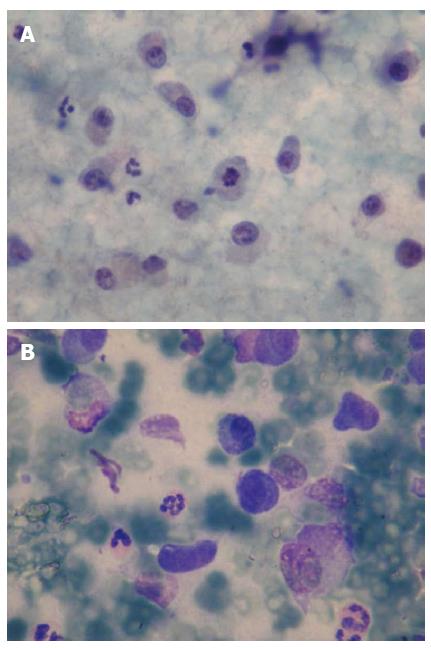
EUS (Fujinon, Tokyo, Japan) revealed a 3 cm heterogeneous focal mass in the head of the pancreas (Figure 1). Neoplastic cells were detected by FNA (22 G; Cook Endoscopy, Winston-Salem, NC, United States), and plasmacytoma was diagnosed by the cytopathologist (Figure 2). Since plasmacytoma features are nonspecific on EUS and resemble other neoplasms including adenocarcinoma, plasmacytoma should be included in the differential diagnosis of a pancreatic mass, especially in advanced stage multiple myeloma patients. EUS-FNA is a fast and reliable technique for the diagnosis of plasmacytoma.
A 64-year-old man who was referred to our endoscopy unit.
Endosonographic (EUS) fine needle aspiration (FNA) for a pancreatic mass.
Plasmacytoma features are nonspecific on EUS.
EUS revealed a 3 cm heterogeneous focal mass in the head of the pancreas. Neoplastic cells were detected by FNA, and plasmacytoma was diagnosed by the cytopathologist.
Since plasmacytoma features are nonspecific on EUS and resemble other neoplasms including adenocarcinoma, plasmacytoma should be included in the differential diagnosis of a pancreatic mass, especially in advanced stage multiple myeloma patients.
An uncommon manifestation of multiple myeloma is extramedullary plasmacytoma. It is generally localized to nasal fossa and rarely involves the pancreas.
EUS-FNA is a fast and reliable technique for the diagnosis of plasmacytoma.
As mentioned in this study, extramedullary plasmacytoma is a rare presentation of multiple myeloma. So it had innovative significance for this study to report a pancreatic plasmacytoma diagnosed by EUS-FNA.
P- Reviewers: Eysselein VE, Lin MS S- Editor: Ma YJ L- Editor: A E- Editor: Zhang DN
| 1. | Lopes da Silva R. Pancreatic involvement by plasma cell neoplasms. J Gastrointest Cancer. 2012;43:157-167. |


