Published online Apr 16, 2011. doi: 10.4253/wjge.v3.i4.78
Revised: December 19, 2010
Accepted: December 26, 2010
Published online: April 16, 2011
Accidentally swallowed foreign objects are not uncommon but difficult to manage without complications. We describe the case of a 68 year old man who accidentally a swallowed sharp-pointed dental reamer that had reached deep in his jejunum. Double balloon enteroscopic retrieval was performed with polypectomy snare but the reamer was entangled in the wire loop of the snare and penetrated the jejunal wall. After releasing the reamer by pushing and pulling the snare for approximately 30 min, the reamer was retrieved with biopsy forceps. This is the first report of double balloon enteroscopic removal of a dental reamer. Furthermore, this is a novel case with regard to decision making in situations when sharp objects are swallowed.
- Citation: Kato S, Kani K, Takabayashi H, Yamamoto R, Yakabi K. Double balloon enteroscopy to retrieve an accidentally swallowed dental reamer deep in the jejunum. World J Gastrointest Endosc 2011; 3(4): 78-80
- URL: https://www.wjgnet.com/1948-5190/full/v3/i4/78.htm
- DOI: https://dx.doi.org/10.4253/wjge.v3.i4.78
Accidental swallowing of foreign objects is not uncommon and may require emergency medical assistance. However, endoscopic removal of swallowed objects from the small intestine without complications is challenging due to the narrow lumen and long length of this organ. Here we report on the use of double balloon enteroscopy to retrieve an accidentally swallowed dental reamer deep in the jejunum.
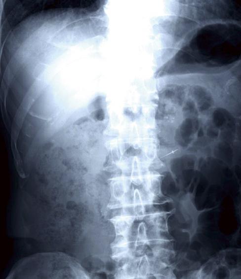
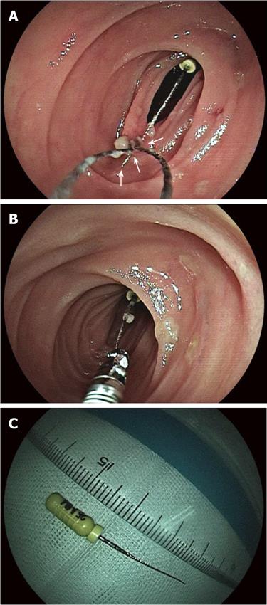
A 68 year old man was admitted to our hospital for accidental swallowing of a dental reamer. On admission, he showed no abnormality in his laboratory data and general well-being. In the past, he had an operation on his tongue due to cancer. Abdominal radiography and CT scanning revealed a dental reamer in his jejunum and no perforation of the gastrointestinal (GI) wall (Figure 1). Double balloon enteroscopic examination was carried out with anterograde approach and the reamer was located deep in the jejunum. Endoscopic removal of the reamer was attempted using a polypectomy snare. Initially, we tried to hold the tip of the reamer but the reamer became entangled in the wire loop of the snare. The patient spontaneously belched, the endoscope was pulled out to the oral side and the needle of the reamer which was entangled in the snare wire penetrated the jejunal wall (Figure 2A). We tried to free the reamer by pushing and pulling the snare but the reamer did not come off the jejunal wall because the flexible arm of the snare only stretched or bent over. Instead, we tried to push and pull the scope backward towards the oral side, hoping to avoid bending the snare arm. Finally, we released the reamer from the jejunal wall in approximately 30 min. After release of the reamer, we thoroughly examined the penetration site. There was no mucosal lesion. Next, we managed to hole the tip of the reamer needle by using biopsy forceps and removed it from the jejunum without further complications (Figure 2B, C).
The majority of foreign bodies which enter the GI tract pass through harmlessly with the feces. The literature shows that 10%-20% of these events require non-surgical intervention and approximately 1% requires surgery[1-3]. However, the guideline for the management of ingested foreign bodies from the American Society for Gastrointestinal Endoscopy indicates that the complications caused by ingestion of sharp-pointed objects are as high as 35%[4]. In the case of the small intestine, complications associated with sharp-pointed objects include ingestion of fish bones[5], pins[6], toothbrush[7], toothpicks[8] and needle[9].
To the best of our knowledge, this is the first report of double balloon enteroscopic removal of a foreign body (dental reamer) from the small intestine. Previously, only one publication in the English language has reported colonoscopic removal of a dental reamer from the colon[10]. In the aforementioned case, the reamer was retrieved by passing a polypectomy snare around the sharp end of the reamer and then the colonoscope was gently withdrawn with the reamer sharp end trailing to avoid injury. In the present case, it took some time to release the reamer from the jejunal wall due to the narrow lumen and long length of the jejunum; contact between the sharp end of the reamer and the jejunal wall was difficult to avoid. Additionally, there was restricted movement and difficulty in manipulating the enteroscope because it had reached deep into the jejunum. Finally, the driving force could not be transmitted to the snare due to the flexibility of the snare arm. We think that enteroscopic removal of foreign objects from the small intestine demands skilful handling of endoscopic instruments as dictated by the long length and narrow lumen of the small intestine. We retrieved a sharp foreign object by a combination of chance and skill. However, this operation was complicated by the anatomy of the organ and carried a high risk of perforation. Another option might have been to retrieve the dental reamer through the overtube together with the endoscope[11]. Another choice is single balloon enteroscopy but this was not available to us in this situation. Single balloon enteroscopy is easy to push and pull the fiberscope through the overtube and the jejunal wall can be protected by pulling sharp-pointed objects into the overtube. Accordingly, our impression is that enteroscopical retrieval of sharp objects should be via the over-tube. Finally, this is a novel case with regard to decision making in a situation when sharp objects are swallowed.
Peer reviewers: Shuji Yamamoto, MD, Department of Gastroenterology and Hepatology, Graduate School of Medicine, Kyoto University, 54 Shogoin Kawahara-cho, Sakyo-ku, Kyoto 606-8507, Japan; Kinichi Hotta, MD, Department of Gastroenterology, Saku Central Hospital, 197 Usuda, Saku, Nagano 384-0301, Japan
S- Editor Zhang HN L- Editor Roemmele A E- Editor Liu N
| 1. | Webb WA. Management of foreign bodies of the upper gastrointestinal tract: update. Gastrointest Endosc. 1995;41:39-51. |
| 3. | Vizcarrondo FJ, Brady PG, Nord HJ. Foreign bodies of the upper gastrointestinal tract. Gastrointest Endosc. 1983;29:208-210. |
| 4. | Eisen GM, Baron TH, Dominitz JA, Faigel DO, Goldstein JL, Johanson JF, Mallery JS, Raddawi HM, Vargo JJ 2nd, Waring JP. Guideline for the management of ingested foreign bodies. Gastrointest Endosc. 2002;55:802-806. |
| 5. | Shibuya T, Osada T, Asaoka D, Mori H, Beppu K, Sakamoto N, Suzuki S, Sai JK, Nagahara A, Otaka M. Double-balloon endoscopy for treatment of long-term abdominal discomfort due to small bowel penetration by an eel bone. Med Sci Monit. 2008;14:CS107-CS109. |
| 6. | Akçam M, Koçkar C, Tola HT, Duman L, Gündüz M. Endoscopic removal of an ingested pin migrated into the liver and affixed by its head to the duodenum. Gastrointest Endosc. 2009;69:382-384. |
| 7. | Lee TH, Lee SH, Park JH, Park do H, Park JY, Park SH, Chung IK, Kim SJ. Endoscopic management of duodenal perforation secondary to ingestion of an uncommon foreign body. Gastrointest Endosc. 2008;67:729-731; discussion 731. |
| 8. | Wichmann MW, Huttl TP, Billing A, Jauch KW. Laparoscopic management of a small bowel perforation caused by a toothpick. Surg Endosc. 2004;18:717-718. |
| 9. | Duseja A, Sachdev A, Malhotra HS. Endoscopic removal of needles from duodenum. Indian J Gastroenterol. 1999;18:91. |
| 10. | Thomas HF, Carr-Locke D. The endoscopic removal of a dental reamer from the colon. Dent Update. 1989;16:214-215. |
| 11. | Yano T, Yamamoto H. Current state of double balloon endoscopy: the latest approach to small intestinal diseases. J Gastroenterol Hepatol. 2009;24:185-192. |


