Published online Nov 16, 2011. doi: 10.4253/wjge.v3.i11.225
Revised: July 20, 2011
Accepted: October 15, 2011
Published online: November 16, 2011
The treatment of choice for patients with unresectable neoplastic obstruction of the small intestine is the placement of expandable metal stents. However, endoscopic delivery from the distal duodenum can be more difficult. This case, shows the usefulness and technical advantages of the overtube and single balloon enteroscopy in the treatment of neoplastic stenosis affecting the small intestine.
- Citation: Espinel J, Pinedo E. A simplified method for stent placement in the distal duodenum: Enteroscopy overtube. World J Gastrointest Endosc 2011; 3(11): 225-227
- URL: https://www.wjgnet.com/1948-5190/full/v3/i11/225.htm
- DOI: https://dx.doi.org/10.4253/wjge.v3.i11.225
Obstruction of the small intestine may be due to malignant neoplasms of adjacent organs. In neoplastic stenosis affecting the gastric exit and the proximal duodenum, palliative treatment with self-expanding metallic stents employing endoscopes of large working channel is a widely used and generally simple option[1,2]. However, when the neoplastic stenosis is more distal (distal duodenum), serious technical limitations related to the endoscopes and the stent make placement challenging.
We present a case of malignant neoplastic obstruction of the distal duodenum-angle of treitz, treated palliatively with placement of an enteral stent, using a single balloon enteroscope.
The patients was a 73-year-old man with clinical signs of intestinal obstruction. An abdominal computed tomography scan revealed inoperable neoplasm of the pancreas infiltrating the third duodenal section (angle of Treitz). Placement of a palliative enteral stent was requested.
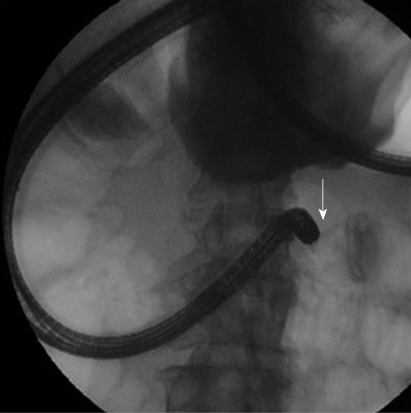
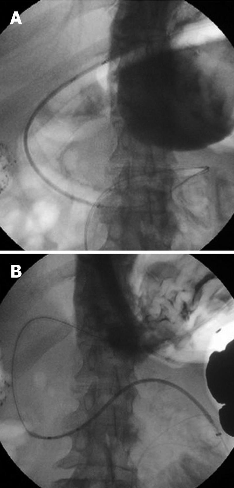
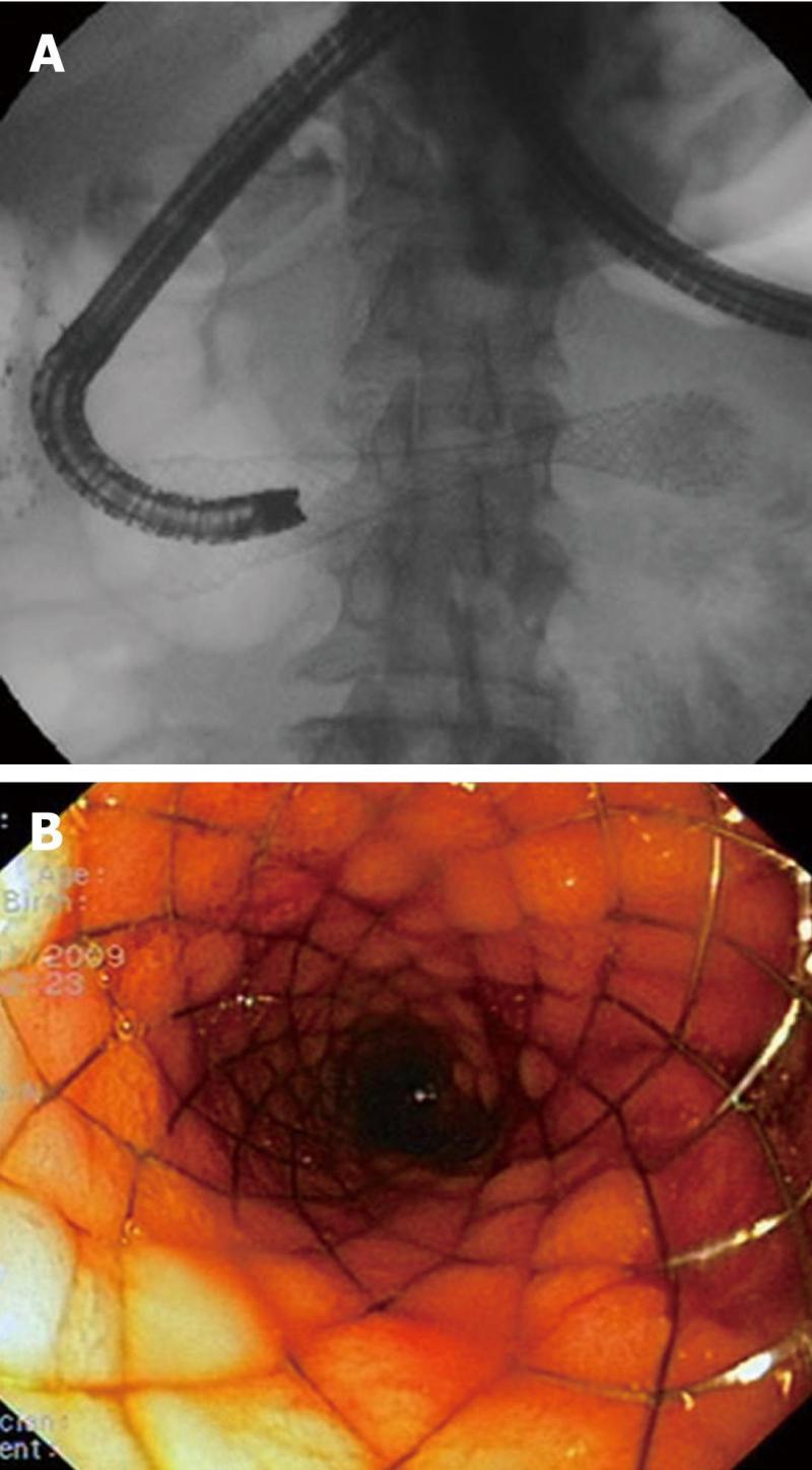
With the patient under general anaesthesia, the stenosis was accessed with a single balloon enteroscope (Olympus SIF Q180) with overtube (ST-SB1). A contrast medium was injected in order to delimit the stenosis (Figure 1). A 0.35-mm guide (Jagwire®, Boston sc) was passed through the stenosis leaving the overtube (OT), and the enteroscope was then withdrawn. Under fluoroscopic control, an enteral stent (22 mm × 90 mm - Wallflex®, Boston) was advanced over the guide and through the OT until it passed through the stenosis (Figure 2A and B). Then, the OT was removed and the stent was released in the stenosis (Figure 3A and B).
The patient tolerated a liquid diet after 24 h, and was subsequently discharged.
The OT is a semi-rigid plastic device designed to facilitate the performance of endoscopy. Its purpose is to protect the gastrointestinal mucosa from trauma and reduce the risk of aspiration. It also facilitates access in patients with difficult anatomy, increases the depth of insertion and maintains access for repeated entry and withdrawal of the endoscope. Potential indications for the use of an OT during upper digestive tract endoscopy are: extraction of foreign bodies orally (prevention of damage to the oesophageal mucosa and aspiration), facilitation of endoscopic intubation (mucosal resection, variceal ligation), protection of altered anatomy (Zenker’s diverticulum), the incorporation of specialized endoscopies (single or double balloon enteroscope, therapeutic endoscopic retrograde cholangiopancreatography in patients with surgically altered anatomy, for cholangioscopy), reduction of infection or neoplastic seeding during placement of a percutaneous endoscopic gastrostomy. In the case of enteroscopy, use of OT can to reduce looping, allowing the most distal sections of the intestine to be reached[3]. To our knowledge, use of the OT in the placement of enteral stents is exceptionally well received[4,5] and this is the first detailed case of the use of an OT with simple balloon enteroscopy for this indication. There is interest in the technique in that the OT permits access to a constricted section of the distal duodenum, and the enteral stent can be rapidly and easily released under fluoroscopic control.
Peer reviewers: Ka Ho Lok, MBChB, MRCP, FHKCP, FHKAM, Associate Consultant, Department of Medicine and Geriatrics, Tuen Mun Hospital, Tsing Chung Koon Road, Tuen Mun, Hong Kong, China; Shuji Yamamoto, MD, Department of Gastroenterology and Hepatology, Graduate School of Medicine, Kyoto University, 54 Shogoin Kawahara-cho, Sakyo-ku, Kyoto 606-8507, Japan
S- Editor Zhang SJ L- Editor Hughes D E- Editor Zheng XM
| 1. | Espinel J, Vivas S, Muñoz F, Jorquera F, Olcoz JL. Palliative treatment of malignant obstruction of gastric outlet using an endoscopically placed enteral Wallstent. Dig Dis Sci. 2001;46:2322-2324. [PubMed] |
| 2. | Espinel J, Sanz O, Vivas S, Jorquera F, Muñoz F, Olcoz JL, Pinedo E. Malignant gastrointestinal obstruction: endoscopic stenting versus surgical palliation. Surg Endosc. 2006;20:1083-1087. [PubMed] |
| 3. | Tierney WM, Adler DG, Conway JD, Diehl DL, Farraye FA, Kantsevoy SV, Kaul V, Kethu SR, Kwon RS, Mamula P. Overtube use in gastrointestinal endoscopy. Gastrointest Endosc. 2009;70:828-834. [PubMed] |
| 4. | Samalin E, Assenat E, Bauret P, Senesse P. Self-expandable metal stents placed with overtube for the treatment of malignant obstruction of the gastrointestinal tract in 33 consecutive patients. Endoscopy. 2007;39 Suppl 1:E101. [PubMed] |
| 5. | Ross AS, Semrad C, Waxman I, Dye C. Enteral stent placement by double balloon enteroscopy for palliation of malignant small bowel obstruction. Gastrointest Endosc. 2006;64:835-837. [PubMed] |


