Published online Jan 16, 2011. doi: 10.4253/wjge.v3.i1.11
Revised: December 12, 2010
Accepted: December 19, 2010
Published online: January 16, 2011
AIM: To conduct a preliminary study on the effect of flexible spectral imaging color enhancement (FICE) used in combination with ultraslim endoscopy by focusing on the enhanced contrast between tumor and non-tumor lesions.
METHODS: We examined 50 lesions of 40 patients with epithelial tumors of the upper gastrointestinal tract before endoscopic submucosal dissection using ultraslim endoscopy with conventional natural color imaging and with FICE imaging. We retrospectively investigated the effect of the use of FICE on endoscopic diagnosis in comparison with normal light.
RESULTS: Visibility of the epithelial tumors of the upper gastrointestinal tract with FICE was superior to normal light in 54% of the observations and comparable to normal light in 46% of the observations. There was no lesion for which visibility with FICE was inferior to that with normal light. FICE visualized 69.6% of hyperemic lesions and 58.8% of discolored lesions better than conventional endoscopy with natural color imaging. FICE significantly improved the visibility of lesions with hyperemia or discoloration compared with normocolored lesions.
CONCLUSION: This study suggests that the use of FICE would improve the ability of ultraslim endoscopy to detect epithelial tumors of the upper gastrointestinal tract.
- Citation: Tanioka Y, Yanai H, Sakaguchi E. Ultraslim endoscopy with flexible spectral imaging color enhancement for upper gastrointestinal neoplasms. World J Gastrointest Endosc 2011; 3(1): 11-15
- URL: https://www.wjgnet.com/1948-5190/full/v3/i1/11.htm
- DOI: https://dx.doi.org/10.4253/wjge.v3.i1.11
Recent practical application of ultraslim endoscopy has led to the rapid spread of transnasal endoscopy as a screening method that causes less pain than peroral endoscopy for the patient during esophago-gastro-duodenoscopy (EGD)[1,2]. Garcia et al[3] reported that ultraslim transnasal endoscopy required a significantly shorter recovery time with significantly lower costs for recovery rooms, personnel expenses, intravenous access devices and oxygen monitors compared to conventional peroral endoscopy with a sedated patient. In addition, transnasal ultraslim endoscopy is appropriate for patients with trismus, those who cannot accept insertion for peroral endoscopy without sedation, and patients with stenosis of the upper gastrointestinal tract in the pharynx or lower regions[4].
However, there are some disadvantages caused by the downsizing of scopes. Compared with conventional endoscopy, ultraslim endoscopy provides less resolution due to the smaller number of pixels and less illumination. Lower power for supplying air and water necessitates a longer time for yielding good images and the smaller diameters of forceps channels limit the usable instruments, resulting in smaller specimens and less information from tissues from a biopsy. For these reasons, ultraslim endoscopy is mainly used for screening purposes rather than detailed close examination. Thus, the ability of ultraslim endoscopy for detection and early diagnosis of gastric cancer should be studied in detail.
Flexible spectral imaging color enhancement (FICE) is one of the diagnostic methods using specific light spectra based on spectral image processing technology (Fujinon Corporation, Saitama, Japan). Currently, it is mainly employed for detailed diagnostic workups for superficial lesions in the gastrointestinal tract using high-resolution magnifying endoscopy and is gradually spreading. Visible light consists of wavelengths from red to purple. A spectral image, the image captured by each wavelength, of a specific wavelength is electrically amplified and reconstructed to create a FICE image. FICE provides comparison of spectral images of diseased and surrounding normal areas for enhancement of the contrast by combining wavelengths with greater differences in signals[5,6].
In the present study, we focused on the enhanced color contrast between tumor and non-tumor areas in FICE images and conducted a preliminary retrospective study on the effect of FICE used in combination with ultraslim endoscopy for observation of superficial epithelial tumors of the upper gastrointestinal tract.
Our institution introduced the ultraslim endoscopy system for EGD in April 2007 and had examined 380 patients, mainly for screening, by February 2008. During this period, we examined 53 lesions in 42 patients with epithelial tumors of the upper gastrointestinal tract who underwent endoscopic submucosal dissection (ESD). The examination was conducted using ultraslim endoscopy with conventional natural color imaging and with FICE imaging before ESD, after obtaining the consent of the patient. Based on the observation of these 53 lesions, we retrospectively investigated the effect of the use of FICE on the visibility of upper gastrointestinal tumorous lesions. The lesions consisted of 3 superficial carcinomas of the esophagus, 3 gastric non neoplastic polyps, 19 gastric adenomas and 28 early gastric cancers.
We used the EG-530N2 (tip diameter 5.9 mm, forceps channel diameter 2.0 mm, four-way bending; Fujinon Corporation, Saitama, Japan) for ultraslim endoscopy of the upper gastrointestinal tract. For FICE imaging, we selected a wavelength set of 525 nm (4), 495 nm (5) and 495 nm (4) for R, G and B respectively, which provides optimal illumination and highlights hyperemic changes commonly seen in epithelial tumors.
We used a five-point scale (Table 1) to evaluate the visibility of lesions which is related to the ability of screening to detect lesions, based on comparison of conventional images and FICE images mainly in long- and middle-distance views.
| Point | Evaluation of observation with FICE |
| 1 | FICE fails to visualize lesions detectable with conventional images |
| 2 | FICE is slightly inferior to conventional images |
| 3 | FICE is comparable to conventional images |
| 4 | FICE is superior to conventional images |
| 5 | FICE allows detection of lesions not easily detected with conventional images or clearer visualization of areas poorly defined with conventional images |
In addition, the lesions were classified as discolored, hyperemic or normocolored (no color differences from surrounding mucosa) for comparison of visibility in the observation with FICE. Fisher’s exact probability test was used for statistical analysis.
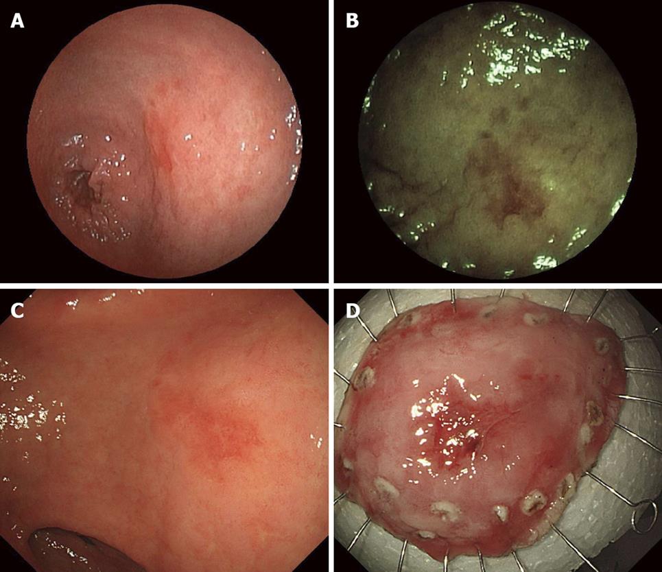
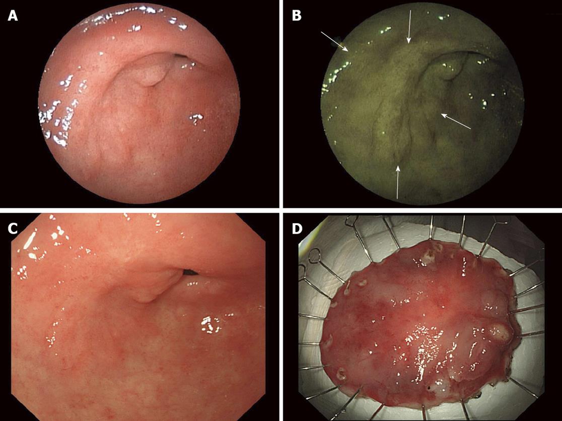
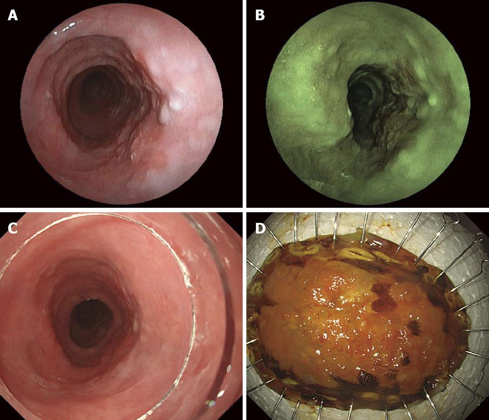
No lesions were graded 1 or 2, inferior to conventional images, for visibility with FICE. In 24 of the 53 lesions (45.3%), visibility with FICE was graded 3, comparable to conventional images. In 29 of the 53 lesions (54.7%), visibility with FICE was graded 4, superior to conventional images (Table 2, Figures 1, 2 and 3).
| Evaluation of FICE | 1 | 2 | 3 | 4 | 5 | Subtotal |
| Superficial carcinoma of the esophagus | 0 | 0 | 2 | 1 | 0 | 3 |
| Gastric non neoplastic polyp | 0 | 0 | 3 | 0 | 0 | 3 |
| Gastric adenoma | 0 | 0 | 8 | 11 | 0 | 19 |
| Early gastric cancer | 0 | 0 | 11 | 17 | 0 | 28 |
| 0 | 0 | 24 | 29 | 0 | 53 |
Regarding color hue changes in the lesions, 17 of 25 lesions (68.0%) with hyperemic changes were better visualized with FICE than conventional images (FICE 4, Figures 1-3). Eleven of 18 discolored lesions (61.1%) were also better visualized with FICE. However, nine of ten normocolored lesions (90%) detected with conventional images were similarly visualized with FICE (Table 3). In short, FICE significantly improved the visibility of lesions with hyperemia (P = 0.0027) or discoloration (P = 0.0159) compared with normocolored lesions.
To aid examination and diagnosis using conventional endoscopy, conventional chromoendoscopy with the spraying of a dye such as indigo carmine highlights the surface irregularities of lesions and is a common and very useful method for defining lesions. However, there are problems: additional costs for spraying the dye, the time involved and the inability to highlight the capillary patterns which is important for the early diagnosis of cancer[7].
Virtual chromoendoscopy systems were developed to correct these problems[5]. FICE uses spectral estimation technology to pick up different given wavelengths from all wavelength components from the CCD and produces images using arithmetical processing. FICE provides real-time switching at the flick of a switch. In addition, as FICE electrically amplifies spectral images of given wavelengths and reconstructs the images, it offers brighter images with subtle highlighted color changes and hyperemic areas on the surface of the mucosa[8]. Therefore, we obtained sufficient illumination for long-distance observation with FICE in all cases in this study.
FICE observation of superficial epithelial tumors of the gastrointestinal tract resulted in superior visibility for 54.7% and comparable visibility for 45.3% compared with conventional images. These results suggested that FICE would be useful as a diagnostic aid for ultraslim endoscopy which is becoming more common. FICE significantly improved the visibility of lesions with hyperemia (68.0%) and discoloration (61.1%) compared with conventional images. The results showed that, due to the characteristics of FICE, observation by ultraslim endoscopy with FICE more clearly visualized the color contrast between diseased and normal lesions than observation with conventional images. FICE was also expected to improve the visibility of lesions with color contrast in the mucosa in long-distance observation by lower-resolution ultraslim endoscopy.
Yoshida reported that a review of endoscopic observation of gastritis-like early gastric cancer diagnosed as benign by conventional endoscopy revealed discolored lesions in 29 of 132 cases (22.0%), hyperemic lesions in 83 cases (62.9%) and normocolored lesions in 20 cases (15.2%)[7]. These lesions, not easily diagnosed by conventional endoscopy, may be overlooked in observations with conventional ultraslim endoscopy. Of these lesions, discolored and hyperemic lesions accounted for 84.9%. FICE was considered to prevent overlooking lesions with such color changes.
This study suggests that the use of FICE would improve the ability of ultraslim endoscopy to detect epithelial tumors of the upper gastrointestinal tract under conditions without the spraying of a dye or a biopsy.
The reduction in the endoscope diameter would improve the acceptance of unsedated endoscopy. However, ultraslim endoscopy provides less resolution due to smaller number of pixels and less illumination. Flexible spectral imaging color enhancement (FICE) is one of the diagnostic methods using specific light spectra based on spectral image processing technology (Fujinon Corporation, Saitama, Japan). We focused on the effect of FICE used in combination with ultraslim endoscopy for observation of superficial epithelial tumors of the upper gastrointestinal tract.
The analysis and enhancement of diagnostic accuracy of ultraslim endoscopy is a new frontier for upper gastrointestinal cancer surveillance.
FICE provides comparison of spectral images of diseased and surrounding normal areas for enhancement of the contrast by combining wavelengths with greater differences in signals.
Our study suggests that the use of FICE would improve the ability of ultraslim endoscopy to detect epithelial tumors of the upper gastrointestinal tract under conditions without the spraying of a dye or a biopsy.
Ultraslim endoscopes: a shaft diameter of 6 mm or less which allows them to be passed through the nose or mouth. Flexible spectral Imaging Color Enhancement (FICE): Visible light consists of wavelengths from red to purple. A spectral image, the image captured by each wavelength, of a specific wavelength is electrically amplified and reconstructed to create a FICE image.
Overall, the manuscript is good. The authors show flexible spectral imaging color enhancement could improve ultraslim endoscopy in detection of epithelial tumors of the upper gastrointestinal tract. Therefore I could recommend publication if several points were discussed.
Peer reviewer: Hongchun Bao, PhD, Research Fellow, The Center for Micro-Photonics, Faculty of Engineering & Industrial Sciences, Swinburne University of Technology, PO Box 218, Hawthorn, Victoria 3122, Australia
S- Editor Zhang HN L- Editor Roemmele A E- Editor Liu N
| 1. | Birkner B, Fritz N, Schatke W, Hasford J. A prospective randomized comparison of unsedated ultrathin versus standard esophagogastroduodenoscopy in routine outpatient gastroenterology practice: does it work better through the nose? Endoscopy. 2003;35:647-651. |
| 2. | Preiss C, Charton JP, Schumacher B, Neuhaus H. A randomized trial of unsedated transnasal small-caliber esophagogastroduodenoscopy (EGD) versus peroral small-caliber EGD versus conventional EGD. Endoscopy. 2003;35:641-645. |
| 3. | Garcia RT, Cello JP, Nguyen MH, Rogers SJ, Rodas A, Trinh HN. Unsedated ultrathin EGD is well accepted compared with conventional sedated EGD: a multicenter randomized trial. Gastroenterology. 2003;125:1606-1612. |
| 4. | Abe K, Miyaoka M. Trial of transnasal esophagogastroduodenoscopy. Digestive Endoscopy. 2006;18:212-217. |
| 5. | Pohl J, May A, Rabenstein T, Pech O, Ell C. Computed virtual chromoendoscopy: a new tool for enhancing tissue surface structures. Endoscopy. 2007;39:80-83. |
| 6. | Osawa H, Yoshizawa M, Yamamoto H, Kita H, Satoh K, Ohnishi H. Optimal band imaging system can facilitate detection of changes in depressed-type early gastric cancer. Gastrointest Endosc. 2008;67:226-234. |
| 7. | Yoshida S. Endoscopy, Gastric Cancer. Oxford University Press: Tokyo 1997; 168-188. |
| 8. | Mouri R, Yoshida S, Tanaka S, Oka S, Yoshihara M, Chayama K. Evaluation and validation of computed virtual chromoendoscopy in early gastric cancer. Gastrointestinal Endoscopy. 2009;69:1052-1058. |


