Case Report Open Access
Copyright ©2010 Baishideng. All rights reserved.
World J Gastrointest Endosc. May 16, 2010; 2(5): 186-189
Published online May 16, 2010. doi: 10.4253/wjge.v2.i5.186
Endoscopic removal of multiple duodenum foreign bodies: An unusual occurrence
Sameer R Islam, Ebtesam A Islam, David Hodges, Kenneth Nugent, Sreerum Parupudi, Department of Internal Medicine and Gastroenterology, Texas Tech University Health Sciences Center, 3601 4th Street, Lubbock, TX 79430, United States
Author contributions: Islam SR, Islam EA, Nugent K, Parupudi S, Hodges D contributed equally to this work; Islam SR, Nugent K, Parupudi S obtained permission from the Texas Department of Criminal Justice to publish this work; Islam SR, Nugent K, Parupudi S contributed in writing the manuscript; and all authors have read and approved the paper.
Correspondence to: Sameer Islam, MD, Department of Internal Medicine and Gastroenterology, Texas Tech University Health Sciences Center, 3601 4th Street, Lubbock, TX 79430, United States. sameer.islam@ttuhsc.edu
Telephone: +1-806-7433155 Fax: +1-806-7433148
Received: November 10, 2009
Revised: March 26, 2010
Accepted: April 2, 2010
Published online: May 16, 2010

Abstract

Deliberate single foreign body ingestion is a scenario that many gastroenterologists commonly see in psychiatric units and prisons. However, multiple foreign body ingestions, especially located in the duodenum, provide the endoscopist with unique challenges for management and treatment. Although most foreign objects pass spontaneously, one should have a low threshold of intervention for multiple objects, especially those that are wide, sharp and at risk of perforation. Diagnosis is typically made when there is a history of ingestion coupled with corresponding radiographic verification. The symptoms tend to be non-specific although some patients are able to delineate where the discomfort level is, correlating with the site of impaction. Most foreign bodies pass spontaneously; however when multiple sharp objects are ingested, the gastroenterologist should perform endoscopic procedures to minimize the risks of bowel perforation. We describe here a successful case of multiple ingested foreign bodies retrieved across the C-loop of the duodenum and the pharynges-esophageal curve via endoscopy and review the literature of multiple foreign body ingestion.

Key Words: Foreign bodies; Multiple; Duodenum; Management; Foreign body ingestions


INTRODUCTION

Foreign body ingestions are a common feature of many patients who are young, alcoholic or have psychiatric conditions and it is a common scenario many primary care physicians or gastroenterologists see. The dilemma is whether to allow the foreign body to pass spontaneously or to remove it either endoscopically or surgically. We report a case of a schizophrenic prisoner with a history of bowel resection due to past foreign body ingestions who came to the emergency room after swallowing multiple spoons and a ballpoint pen. These were successfully retrieved endoscopically. In this report we review the literature on multiple foreign body ingestions located in the duodenum and possible interventions for endoscopic management.

CASE REPORT

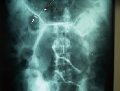
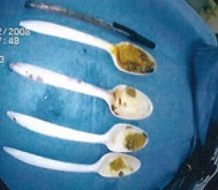
A 62-year-old male prisoner with schizophrenia was brought to the emergency room with severe abdominal pain and recurrent vomiting. He has a previous history of several instances of uneventful foreign body ingestions. However, three years ago one such episode resulted in bowel perforations that eventually required colectomy and end ileostomy. Currently he reported ingestion of multiple plastic spoons and a ballpoint pen three days previously. He had dry oral mucosa, normal vital signs and a mildly distended abdomen with no signs of obstruction or peritonitis. Serial abdominal radiographs showed linear outlines of the ingested foreign bodies in the subhepatic region that have not changed in position (Figure 1). These were presumed to be in the second and third parts of duodenum. He had hypokalemia (2.9 mEq/dL) that was corrected with intravenous fluids and potassium supplementation. An upper endoscopy was performed under monitored anesthesia care. Multiple plastic spoons and a ballpoint pen were impacted in the distal C-loop of duodenum. All the spoons were oriented with their handles directed proximally. Using a polypectomy snare to grasp the distal handle of the most accessible spoon, it was gently disimpacted and brought into the stomach. There the snare was reoriented to align the spoon vertically grasping the distal handle about 1 cm from the tip. Then it was gradually brought out across the GE junction up to the pharyngo-esophageal curve where maneuvering the rigid, non-yielding foreign body was difficult. To accomplish the maneuver, the foreign body was held with the snare as far up in the hypopharynx as possible by the endoscopist while the anesthetist introduced grasping forceps through the mouth aided by a direct laryngoscope. The tip of the spoon was grasped with the forceps and gently maneuvered across the hypopharynx without trauma. Four spoons and a ballpoint pen were successfully retrieved by this method, one after the other (Figure 2). Post-procedure check endoscopy showed no significant mucosal trauma or bleeding and a repeat abdominal x-ray confirmed that all the foreign bodies were removed. The patient was counseled against foreign body ingestion and care was transferred to the psychiatry service.

Figure 1
Figure 1 Abdominal X-ray demonstrating the metal part of the pen (short white arrow) and outlines of multiple plastic spoons (long white arrows).
Figure 2
Figure 2 Successful extraction of all the duodenal foreign bodies.
DISCUSSION

In adults, most foreign body ingestion occurs in certain populations: the elderly, mentally disabled, alcoholics, prisoners and psychiatric patients[1-3]. Most foreign bodies will pass spontaneously through the gastrointestinal tract without any complications[1]; in fact, once past the esophagus the majority of foreign bodies ingested will move uneventfully throughout the alimentary canal[4,5]. However, non-operative interventions are still necessary in 10%-20% of patients and surgery in 1%[1], especially in the cases of ingested multiple sharp and long objects.

Timely diagnosis can be difficult as the ingestion goes unreported until the onset of symptoms which may be remote from the actual time of ingestion[6,7]. Radiograph films can identify most foreign objects; however, they do not readily detect fish or chicken bones, wood, plastic, most glass and thin metals objects[8]. Contrast examinations with barium should not be performed routinely because coating the foreign body and esophageal mucosa can compromise the subsequent endoscopy[9]; they should be performed cautiously if symptoms are not clear in order to clarify the presence of a foreign body[10]. CT scans, though commonly used, can be negative with radiolucent objects though their yield can be enhanced with 3D reconstruction[11,12].

Management decision for patients depend on a variety of factors including age, the object ingested, the location of the ingested object, the urgency of removal, the number of objects swallowed and the skill of the endoscopist. The timing should be contingent on the perceived risks of obstruction and/or perforation. Most physicians would prefer the endoscopic route since it avoids surgery, has reduced costs, is relatively accessible, allows simultaneous diagnosis of other diseases and has a low rate of mortality[3]. Endoscopic removal of foreign objects should be performed by experienced endoscopists using accessories such as snares, Dormia baskets or strong-toothed graspers[13]. Objects longer than 6 to 10 cm such as spoons and toothbrushes should be removed using a longer (> 45 cm) overtube that extends beyond the gastroesophageal junction. The object can be grasped with a snare or basket and maneuvered into the overtube[1]. The entire apparatus, foreign body, overtube and endoscope can then be withdrawn in one motion, avoiding losing grasp of the object in the overtube itself[14]. The endoscopic retrieval of sharp objects is accomplished using retrieval forceps (rat-tooth, biopsy or alligator jaws) or a snare. The risk of mucosal injury during sharp-object retrieval can be minimized by orientating the object with the point trailing during the extraction with an overtube[13]. In the presented case, endoscopic retrieval could have been facilitated by using an overtube but all prison systems are not equipped for advanced endoscopic interventions and lack of it did not deter successful retrieval of the multiple swallowed objects.

Urgent retrieval is necessary to avoid complications of the object ingested. Those patients with a past history of gastrointestinal tract surgery or congenital gut malformations are at an increased risk of obstruction or perforation. Our patient had a past history of gastrointestinal operations, mitigating the possibility of spontaneous evacuation. Additionally, the properties of the objects themselves determine the degree of complications associated with ingestion. Long, slender items have a more difficult time transversing the tortuous gastrointestinal tract; hence they are more likely to stay lodged[4,15]. This is further complicated by ingesting multiple long, slender objects each of which carries the risk of obstruction or perforation. In general, objects wider than 2 cm do not pass though the pylorus and tend to lodge in the stomach while objects longer than 5 cm tend to get caught in the duodenal sweep[16,17]. Additionally, it is recommended that sharp-pointed objects like pens in this case, should be removed even if the patient is asymptomatic[13] as the mortality and the risk of perforations increases with these objects[1,2,5] leading to peritonitis, abscess formation, inflammatory mass formation, obstruction, fistulae, hemorrhage or even death[15,18].

Complicating the clinical picture in this case were the multiple foreign bodies lodged in the duodenum. In reviewing the literature, several retrospective case series did not delineate a clear approach to multiple foreign bodies obstructed in the duodenum. One series involving a retrospective analysis of foreign body ingestions in Greece noted only 2.6% of ingested foreign bodies were lodged in the duodenum[19]; none of these cases involved multiple objects. Similarly a comparison of 1088 cases in China showed that most of the foreign body ingestions were located in the esophagus (53%) while only 4.5% were found in the duodenum[3]. Other series of foreign body ingestions in Italy[20], Bulgaria[15], the United States[21], Korea[22], Jordan[23] and Greece[24] showed similar findings with singular objects ingested. We were able to successfully retrieve multiple objects via endoscopy by manipulating the snare carefully while gently maneuvering the gasping forceps onto the objects, thereby easing the foreign bodies through the hypopharynx individually and avoiding a surgical procedure.

Deliberate ingestion of foreign bodies by prison inmates and psychiatry patients pose unique challenges for endoscopic removal. Though most objects can pass spontaneously, the presentation of symptoms necessitated a treatment option. It is important to evaluate each individual case with respect to the comfort level of the endoscopist and the emergent nature of the intervention. Attempt at endoscopic retrieval is the current standard of care for foreign body ingestions in the absence of features of perforation or major vessel penetration. Impaction of multiple long, rigid objects in the duodenum and their successful retrieval across the C-loop of the duodenum and the pharynges-esophageal curve are the unique features in this reported case.

Footnotes

Peer reviewers: Alfredo José Lucendo, MD, PhD, Department of Gastroenterology, Hospital General de Tomelloso, Vereda de Socuéllamos, s/n, Tomelloso (Ciudad Real) 13700, Spain; Sherman M Chamberlain, MD, FACP, FACG, AGAF, Associate Professor of Medicine, Section of Gastroenterology, BBR-2538, Medical College of Georgia, Augusta, GA 30912, United States

S- Editor Zhang HN L- Editor Roemmele A E- Editor Liu N

References
1.  Eisen GM, Baron TH, Dominitz JA, Faigel DO, Goldstein JL, Johanson JF, Mallery JS, Raddawi HM, Vargo JJ 2nd, Waring JP. Guideline for the management of ingested foreign bodies. Gastrointest Endosc. 2002;55:802-806.  [PubMed]  [DOI]  [Cited in This Article: ]
2.  Stiles BM, Wilson WH, Bridges MA, Choudhury A, Rivera-Arias J, Nguyen DB, Edlich RF. Denture esophageal impaction refractory to endoscopic removal in a psychiatric patient. J Emerg Med. 2000;18:323-326.  [PubMed]  [DOI]  [Cited in This Article: ]
3.  Li ZS, Sun ZX, Zou DW, Xu GM, Wu RP, Liao Z. Endoscopic management of foreign bodies in the upper-GI tract: experience with 1088 cases in China. Gastrointest Endosc. 2006;64:485-492.  [PubMed]  [DOI]  [Cited in This Article: ]
4.  Webb WA. Management of foreign bodies of the upper gastrointestinal tract: update. Gastrointest Endosc. 1995;41:39-51.  [PubMed]  [DOI]  [Cited in This Article: ]
5.  Vizcarrondo FJ, Brady PG, Nord HJ. Foreign bodies of the upper gastrointestinal tract. Gastrointest Endosc. 1983;29:208-210.  [PubMed]  [DOI]  [Cited in This Article: ]
6.  Adams DB. Endoscopic removal of entrapped coins from an intraluminal duodenal diverticulum 20 years after ingestion. Gastrointest Endosc. 1986;32:415-416.  [PubMed]  [DOI]  [Cited in This Article: ]
7.  Tsui BC, Mossey J. Occult liver abscess following clinically unsuspected ingestion of foreign bodies. Can J Gastroenterol. 1997;11:445-448.  [PubMed]  [DOI]  [Cited in This Article: ]
8.  Cheng W, Tam PK. Foreign-body ingestion in children: experience with 1,265 cases. J Pediatr Surg. 1999;34:1472-1476.  [PubMed]  [DOI]  [Cited in This Article: ]
9.  Ginsberg GG. Management of ingested foreign objects and food bolus impactions. Gastrointest Endosc. 1995;41:33-38.  [PubMed]  [DOI]  [Cited in This Article: ]
10.  Seikel K, Primm PA, Elizondo BJ, Remley KL. Handheld metal detector localization of ingested metallic foreign bodies: accurate in any hands? Arch Pediatr Adolesc Med. 1999;153:853-857.  [PubMed]  [DOI]  [Cited in This Article: ]
11.  Cranston PE, Pollack CV Jr, Harrison RB. CT of crack cocaine ingestion. J Comput Assist Tomogr. 1992;16:560-563.  [PubMed]  [DOI]  [Cited in This Article: ]
12.  Takada M, Kashiwagi R, Sakane M, Tabata F, Kuroda Y. 3D-CT diagnosis for ingested foreign bodies. Am J Emerg Med. 2000;18:192-193.  [PubMed]  [DOI]  [Cited in This Article: ]
13.  Cho HJ, Kim JY, Lee HC, Kweon YO, Cho CM, Tak WY, Jeon SW. An impacted clamshell in the duodenum mistaken for a gall stone. Korean J Intern Med. 2007;22:292-295.  [PubMed]  [DOI]  [Cited in This Article: ]
14.  Chinitz MA, Bertrand G. Endoscopic removal of toothbrushes. Gastrointest Endosc. 1990;36:527-530.  [PubMed]  [DOI]  [Cited in This Article: ]
15.  Velitchkov NG, Grigorov GI, Losanoff JE, Kjossev KT. Ingested foreign bodies of the gastrointestinal tract: retrospective analysis of 542 cases. World J Surg. 1996;20:1001-1005.  [PubMed]  [DOI]  [Cited in This Article: ]
16.  Webb WA. Management of foreign bodies of the upper gastrointestinal tract. Gastroenterology. 1988;94:204-216.  [PubMed]  [DOI]  [Cited in This Article: ]
17.  Chang JJ, Yen CL. Endoscopic retrieval of multiple fragmented gastric bamboo chopsticks by using a flexible overtube. World J Gastroenterol. 2004;10:769-770.  [PubMed]  [DOI]  [Cited in This Article: ]
18.  Selivanov V, Sheldon GF, Cello JP, Crass RA. Management of foreign body ingestion. Ann Surg. 1984;199:187-191.  [PubMed]  [DOI]  [Cited in This Article: ]
19.  Katsinelos P, Kountouras J, Paroutoglou G, Zavos C, Mimidis K, Chatzimavroudis G. Endoscopic techniques and management of foreign body ingestion and food bolus impaction in the upper gastrointestinal tract: a retrospective analysis of 139 cases. J Clin Gastroenterol. 2006;40:784-789.  [PubMed]  [DOI]  [Cited in This Article: ]
20.  Mosca S, Manes G, Martino R, Amitrano L, Bottino V, Bove A, Camera A, De Nucci C, Di Costanzo G, Guardascione M. Endoscopic management of foreign bodies in the upper gastrointestinal tract: report on a series of 414 adult patients. Endoscopy. 2001;33:692-696.  [PubMed]  [DOI]  [Cited in This Article: ]
21.  Weiland ST, Schurr MJ. Conservative management of ingested foreign bodies. J Gastrointest Surg. 2002;6:496-500.  [PubMed]  [DOI]  [Cited in This Article: ]
22.  Kim JK, Kim SS, Kim JI, Kim SW, Yang YS, Cho SH, Lee BS, Han NI, Han SW, Chung IS. Management of foreign bodies in the gastrointestinal tract: an analysis of 104 cases in children. Endoscopy. 1999;31:302-304.  [PubMed]  [DOI]  [Cited in This Article: ]
23.  Mahafza T, Batieha A, Suboh M, Khrais T. Esophageal foreign bodies: a Jordanian experience. Int J Pediatr Otorhinolaryngol. 2002;64:225-227.  [PubMed]  [DOI]  [Cited in This Article: ]
24.  Gorse GJ, Messner RL. Infection control practices in gastrointestinal endoscopy in the United States: a national survey. Gastroenterol Nurs. 1991;14:72-79.  [PubMed]  [DOI]  [Cited in This Article: ]