Published online Nov 16, 2010. doi: 10.4253/wjge.v2.i11.375
Revised: September 10, 2010
Accepted: September 17, 2010
Published online: November 16, 2010
Plastic stent insertion is a treatment option for pancreatic duct stricture with chronic pancreatitis. However, recurrent stricture is a limitation after removing the plastic stent. Self-expandable metal stents have long diameters and patency. A metal stent has become an established management option for pancreatic duct stricture caused by malignancy but its use in benign stricture is still controversial. We introduce a young patient who had chronic pancreatitis and underwent several plastic stent insertions due to recurrent pancreatic duct stricture. His symptoms improved after using a fully covered self-expandable metal covered stent and there was no recurrence found at follow-up at the outpatient department.
- Citation: Lee KJ, Kim KJ, Shin DH, Chung JW, Park JY, Bang S, Park SW, Song SY. Placement of a fully covered self-expandable metal stent in a young patient with chronic pancreatitis. World J Gastrointest Endosc 2010; 2(11): 375-378
- URL: https://www.wjgnet.com/1948-5190/full/v2/i11/375.htm
- DOI: https://dx.doi.org/10.4253/wjge.v2.i11.375
The treatment of chronic pancreatitis with pancreatic duct stricture is still challenging. Conventional plastic pancreatic duct (PD) stents are unsatisfactory because of recurrent stricture and pain. Recently, self-expandable metal covered stents (SEMS) have been used to treat strictures of the main pancreatic duct in chronic pancreatitis patients. However, its long-term safety and efficacy are uncertain because of epithelial hyperplasia and migration[1]. Some studies have demonstrated that a fully covered SEMS (FCSEMS) was effective in patients with symptomatic refractory PD stricture[2]. Here, we introduce a young patient with refractory PD stricture caused by chronic pancreatitis; the stricture and pain were effectively treated after placement of FCSEMS.
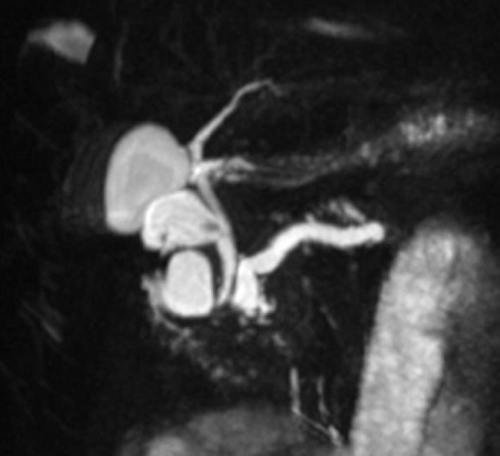
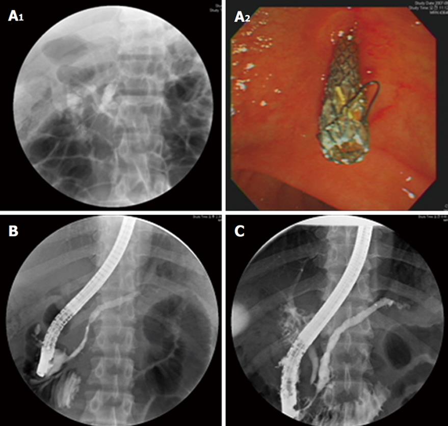
A 13 year old male visited Severance Hospital, Yonsei University College of Medicine with complaints of recurrent abdominal pain. He was diagnosed with chronic pancreatitis at the age of twelve. Magnetic resonance cholangiopancreatography (MRCP) showed chronic pancreatitis and a benign cystic lesion at the pancreatic groove, suggestive of pseudocyst (2.7 cm) (Figure 1). For the treatment of pancreatic pseudocyst, a cysto-duodenal stent (double pigtail, 4 cm) was inserted. One month later, computed tomography (CT) scan showed resolution of the cyst. At three months, the stent had disappeared on CT and no further treatment was provided as he had no symptoms. However, he returned with abdominal pain and vomiting six months after the first trial of the stent. CT showed pancreatic duct dilatation with multiple persistent parenchymal or intraductal calcification, especially at the head of pancreas head, and aggravation of pseudocyst. A plastic PD stent (7 Fr, 5 cm) was inserted and then removed two months later. Another stent of the same size was re-inserted as CT scan showed a remaining cyst. Four months later, pancreatogram showed diffuse dilatation of the pancreatic duct and filling defects at the pancreatic head. A new PD stent with a larger diameter (10 Fr, 5 cm) was inserted. He did not show any symptoms and was admitted to the hospital six months later during his winter break. The CT scan showed resolution of the cyst. However, after removing the stent, pancreatogram showed an irregular stricture and proximal duct dilatation at the main pancreatic duct and pancreatic duct stones were found. Emergent mechanical lithotripsy was used for the treatment of pancreatic stones and the stones were partially removed. He underwent extracorporeal shock wave lithotripsy (ESWL) in the urology department for two days and endoscopic retrograde cholangiopancreatography (ERCP) was done on the day after the last ESWL. The finding still showed PD stones with reduced number and sizes. A plastic PD stent (10 Fr, 5 cm) was inserted for the treatment of pancreatic duct stricture and stones. Four months after inserting the last plastic stent, ERCP showed no definite stricture at the pancreatic duct and the patient was followed up without additional stent insertion. Three months later, he again complained of abdominal pain. ERCP showed that the pancreatic duct was markedly dilated with a stenosis of the head portion but without stones. To use a larger and more expandable device, a removable metal stent (Niti-S type biliary stent, covered, 10 mm, 5 cm, Taewoong, Seoul, Korea) was inserted into the pancreatic duct (Figure 2A). There were no procedure related complications. The patient recovered and was discharged after two days. The follow-up ERCP was performed 1 mo after the stent placement and the stent was successfully removed with an elegator (Figure 2B). After removal of the stent, the pancreatic duct stricture was much improved. Fifteen months after the stent removal, ERCP showed that the patency of the pancreatic duct was maintained (Figure 2C). The patient remains healthy without any signs or symptoms of pancreatitis or need for any pancreatic enzyme supplement or narcotics at 24 mo after stent removal.
About 50% of patients with severe chronic pancreatitis undergoing endoscopic treatment require pancreatic stent placement in order to relieve obstruction of the main pancreatic duct. Even though endoscopic placement of plastic stents has been widely used, it has several limitations[3-5]. For example, since the plastic stents often become occluded with sludge, they usually need to be changed every 3 to 4 mo[6]. Thus, the number of procedures, hospitalization and costs may increase. As an alternative treatment for blockages of plastic stent or refractory stricture after the plastic stent removal, the placement of a metal stent has been considered. Metal stents have a longer patency than polyethylene stents and usually a single metal stent can palliate obstruction for good. The metal stent is effectively used in the treatment of malignant pancreatic duct obstruction. In contrast, its use in patients with benign diseases has yet to be well-established. There have been only a few studies regarding long-term complications, efficacy and outcomes with the use of metal stent in benign disease[7]. In our study, the patient underwent removable metal stent insertion and remained not just free from pain but also had no recurrence of pancreatitis for more than 2 years after the removal of the metal stent. A follow-up pancreatogram also showed resolution of dominant pancreatic ductal strictures. So far, results of self-expandable uncovered metal stents had been disappointing with regard to poor drainage of pancreatic juice and difficult removal due to tissue ingrowth[1,8]. Some studies even recommended that, considering complications such as increasing cancer risk or stent migration, SEMS should not be placed in patients with a benign gastrointestinal disorder, a long life expectancy and who are good candidates for surgery. To overcome these limitations, FCSEMS was developed. A FCSEMS with a larger diameter (up to 6 or 8 mm) may have an advantage in resolving or improving pancreatic ductal stricture compared with a single plastic stent[9]. In addition, the Niti-S type biliary stent (covered, Taewoong) used in this case has a double coated silver membrane which prevents direct contact of the metal with tissue, causes less tissue embedding and can be easily removed. Primary placement of an FCSEMS may, therefore, be an attractive option for refractory benign pancreatic ductal strictures.
Recently, Park et al[9] showed that placement of FCSEMS in patients with refractory benign pancreatic ductal strictures for 2 mo might be feasible and relatively safe. Moon et al[10] reported that temporary 3 mo placement of modified FCSMS with anti-migration features was effective in resolving pancreatic duct strictures in chronic pancreatitis and reduced stent migration. Theoretically, they could provide better drainage and easy removability. However, in their reports, pancreatic sepsis or infection and cholestatic liver dysfunction were still common complications caused by metal stents. Even though FCSEMS seems to be promising, more technological improvement is needed before applying FCSEMS in benign pancreatic diseases in order to be accepted by more endoscopists. Also, further long-term follow-up studies are needed to determine the optimal duration and diameter of FCSEMS for refractory benign pancreatic ductal strictures.
In this case study, a temporary 1 mo placement and removal of a FCSEMS in the main pancreatic duct was deemed feasible and relatively safe. Moreover, the patient had no complications. We expect wider usage of metal stents in benign pancreatic diseases after confirming its long-term efficacy and determining the optimal duration of FCSEMS placement for refractory benign pancreatic ductal strictures through studies with a large number of patients.
Peer reviewers: Rakesh Kumar, Associate Professor, Institute of Liver and Biliary Sciences, New Delhi 110070, India; Peter Born, Professor, Chief, 2nd Department of Medicine, Red Cross Hospital, Munich, Germany; F Douglas Bair, MD, FRCPC, Staff Gastroenterologist, Oakville-Trafalgar Memorial Hospital, Suite 125B - 690 Dorval Drive, Oakville, Ontario L6K 3W7, Canada
S- Editor Zhang HN L- Editor Roemmele A E- Editor Liu N
| 1. | Eisendrath P, Devière J. Expandable metal stents for benign pancreatic duct obstruction. Gastrointest Endosc Clin N Am. 1999;9:547-554. |
| 2. | Kahaleh M, Sundaram V, Condron SL, De La Rue SA, Hall JD, Tokar J, Friel CM, Foley EF, Adams RB, Yeaton P. Temporary placement of covered self-expandable metallic stents in patients with biliary leak: midterm evaluation of a pilot study. Gastrointest Endosc. 2007;66:52-59. |
| 3. | Delhaye M, Arvanitakis M, Bali M, Matos C, Devière J. Endoscopic therapy for chronic pancreatitis. Scand J Surg. 2005;94:143-153. |
| 4. | Eleftherladis N, Dinu F, Delhaye M, Le Moine O, Baize M, Vandermeeren A, Hookey L, Devière J. Long-term outcome after pancreatic stenting in severe chronic pancreatitis. Endoscopy. 2005;37:223-230. |
| 5. | Adler DG, Lichtenstein D, Baron TH, Davila R, Egan JV, Gan SL, Qureshi WA, Rajan E, Shen B, Zuckerman MJ. The role of endoscopy in patients with chronic pancreatitis. Gastrointest Endosc. 2006;63:933-937. |
| 6. | Gilbert DA, DiMarino AJ Jr, Jensen DM, Katon RM, Kimmey MB, Laine LA, MacFaydyen BV, Michaletz-Onody PA, Zuckerman G. Status evaluation: biliary stents. American Society for Gastrointestinal Endoscopy. Technology Assessment Committee. Gastrointest Endosc. 1992;38:750-752. |
| 7. | Sauer B, Talreja J, Ellen K, Ku J, Shami VM, Kahaleh M. Temporary placement of a fully covered self-expandable metal stent in the pancreatic duct for management of symptomatic refractory chronic pancreatitis: preliminary data (with videos). Gastrointest Endosc. 2008;68:1173-1178. |
| 8. | Shin HP, Kim MH, Jung SW, Kim JC, Choi EK, Han J, Lee SS, Seo DW, Lee SK. Endoscopic removal of biliary self-expandable metallic stents: a prospective study. Endoscopy. 2006;38:1250-1255. |
| 9. | Park do H, Kim MH, Moon SH, Lee SS, Seo DW, Lee SK. Feasibility and safety of placement of a newly designed, fully covered self-expandable metal stent for refractory benign pancreatic ductal strictures: a pilot study (with video). Gastrointest Endosc. 2008;68:1182-1189. |
| 10. | Moon SH, Kim MH, Park do H, Song TJ, Eum J, Lee SS, Seo DW, Lee SK. Modified fully covered self-expandable metal stents with antimigration features for benign pancreatic-duct strictures in advanced chronic pancreatitis, with a focus on the safety profile and reducing migration. Gastrointest Endosc. 2010;72:86-91. |


