Published online Oct 16, 2010. doi: 10.4253/wjge.v2.i10.344
Revised: August 31, 2010
Accepted: September 7, 2010
Published online: October 16, 2010
AIM: To evaluate the long-term attachment of two types of endoclips in the human gastrointestinal tract.
METHODS: In this prospective observational study, endoclips were placed and followed-up during endoscopies or using fluoroscopic images as part of a prospective feasibility study evaluating external beam radiotherapy (EBRT, wk 1-3) followed by high dose rate brachytherapy (HDRBT with an endoluminal applicator once a week for 3 wk, wk 9-11) in medically inoperablerectal cancer patients. Initially, the type and number of endoclips were chosenrandomly and later refined to 1 Resolution® clip (Microvasive) proximal and 2 Quickclips® (Olympus) distal to the tumor. Nine consecutive patients from between September 2007 and August 2008 were analyzed. Retention rates were evaluated over three different observational periods [period 1: pre-HDRBT (wk -2-8), period 2: during HDRBT (wk 9-11) and period 3: post-HDRBT (wk 12-16)].
RESULTS: In this study, a total of 44 clips were placed during endoscopy, either at the beginning or at the end of period 1. The Resolution clip had a higher overall retention rate than the Quickclip (P = 0.01). After a median period of 81 d after placement (in period 1), long-term retention rates for the Resolution clip and Quickclip clip were 67% and 35% respectively.
CONCLUSION: The Resolution clip has a high retention rate and is useful in situations where long-term attachment to the human gastrointestinal mucosa is warranted.
- Citation: Swellengrebel HAM, Marijnen CAM, Vincent A, Cats A. Evaluating long-term attachment of two different endoclips in the human gastrointestinal tract. World J Gastrointest Endosc 2010; 2(10): 344-348
- URL: https://www.wjgnet.com/1948-5190/full/v2/i10/344.htm
- DOI: https://dx.doi.org/10.4253/wjge.v2.i10.344
In 1975, Hayashi et al[1] were the first to describe the metallic endoscopic clip as an alternative means to control bleeding by mechanical pressure. Since then, design and clinical indications have been refined. Nowadays, endoscopic clips are frequently used for hemostasis of arterial nonvariceal bleeding of the upper gastrointestinal tract[2,3]. Other reported indications for endoscopic clip placement include the fixation of enteral feeding tubes[4], stent anchorage[5,6] and the management of small fistulas, perforations and anastomotic leaks[7]. Utilizing their radiopaque characteristics, endoclips have recently been used to mark tumors or anatomical structures to facilitate intervention radiology[8], to locate the tumor preoperatively[9] and to delineate tumor volume for radiotherapy[10,11].
Several types of endoscopic clips are commercially available. Most studies involve those from Olympus (Olympus Ltd., Tokyo, Japan), available in preloaded (Quickclip) and reloadable devices (HX-5L). Once the clip has been opened, re-positioning is not possible as the jaws cannot be closed and reopened. The Resolution clip (Microvasive, Boston Scientific Corp, Massachusetts, US) has the ability to reopen its jaws for repositioning which may result in superior positioning and tissue grasping.
The ability of an endoclip to remain attached for a longer period could facilitate procedures in which a tumor needs to be located routinely during the treatment period or when the clip anchors feeding tubes or stents. Retention rates have, however, only been evaluated in canines and pigs and have not yet been reported in the human gastrointestinal tract. The aim of this study was to evaluate the long-term attachment of two endoclips, the Quickclip and the Resolution clip, to human rectal mucosa.
The 9 consecutive patients analyzed in this study were patients with medically inoperable rectal cancer who participated in a prospective feasibility study in the Netherlands Cancer Institute. The primary objective of this ongoing study is to evaluate the feasibility of external beam radiation therapy (EBRT) followed by high-dose rate endorectal brachytherapy (HDRBT) as definitive treatment in patients not suitable for surgery due to co-morbidity, old age or for those refusing surgery. The study gained ethical approval from the Medical Ethics Committee of the Netherlands Cancer Institute. Written informed consent was obtained from all patients. Patient accrual commenced in September 2007.
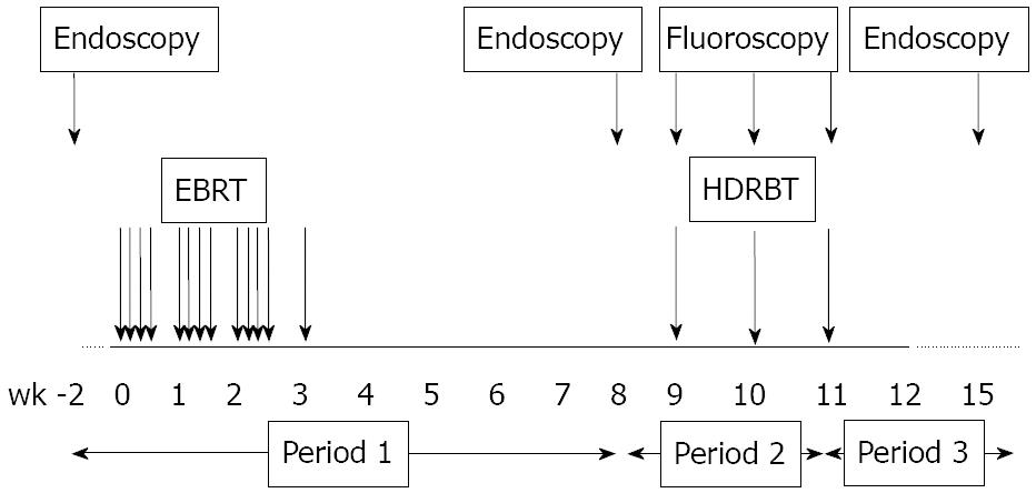
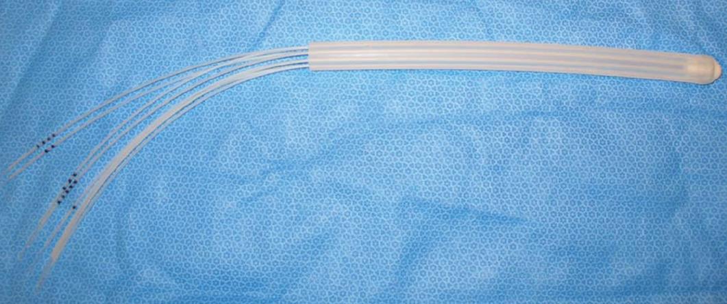
The treatment regimen (Figure 1) consists of 39 Gy administered in 13 fractions of 3 Gy over 3½ wk. After a further 6 wk, HDRBT is applied once every week for 3 wk. HDRBT dose level will be elevated (starting at 5 Gy/fraction) after every 6 patients depending on experienced toxicity. HDRBT is applied using an endorectal applicator (Oncosmart®, Nucletron, Veenendaal, the Netherlands) consisting of a flexible tube with 8 channels (Figure 2). The applicator is 2 cm in diameter and is inserted via the anus prior to each brachytherapy treatment.
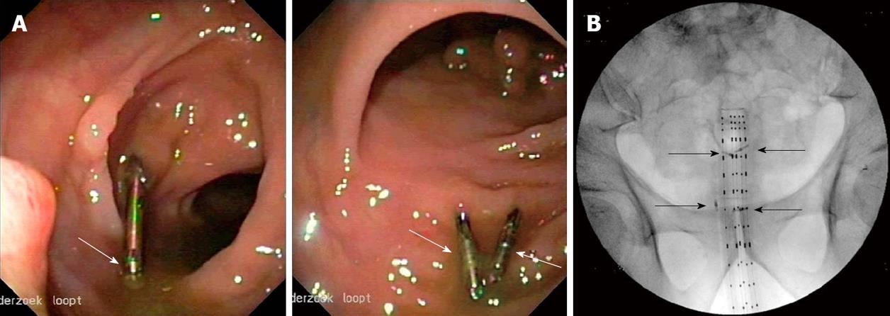
The rectal tumor was marked (Figure 3A) with Quickclips (Olympus Ltd., Tokyo, Japan) and/or Resolution (Microvasive, Boston Scientific Corp, Massachusetts, US) endoclips to facilitate tumor localization during the HDRBT procedure. Endoscopy was performed before EBRT (baseline), before HDRBT (wk 8-9) and after HDRBT (wk 16-17). The tumor was marked before EBRT and, if necessary, additionally (when clips had been dislodged) before HDRBT. Initially, the type and number of endoclips were chosen randomly. Later on, this was refined to one Resolution endoclip at the proximal and two Quickclips at the distal border of the tumor.
A CT-scan with the unloaded applicator inserted was performed for delineation and treatment planning purposes before the first HDRBT fraction. A 3D reconstruction of the applicator and radio-opaque endorectal clips was made and the target volume delineated on the CT-images. A 2D anterior-posterior projection of applicator and clips was reconstructed as a reference for C-arm fluoroscopy guided reinsertion of the applicator prior to each HDRBT session (wk 9-11, Figure 3B).
We evaluated retention rates between the two clip types over three different observational periods [period 1: pre-HDRBT (wk -2-8), period 2: during HDRBT (wk 9-11) and period 3: post-HDRBT (wk 12-16)] (Figure 1). The percentage and absolute number of clips still attached during follow-up endoscopy and on the fluoroscopic images were determined. To assess the retention rates of the two clip types (Quickclip or Resolution) a logistic mixed effects model was constructed with clip type as a fixed effect. To account for the influence of both the different periods (different in length and use of an endoluminal applicator) and the duration of attachment prior to assessment, we included placement (beginning of period 1 or 2) and assessment period as fixed effects. The assessments of clip retention was assumed to be correlated when they relate to the same clip (assessed for different periods) or from clips within the same patient. To account for this cluster correlation, we included clip and patient id as random intercepts. Due to insufficient events in period 2, a logistic model was unable to be constructed; hence the comparison of the retention rates of the two clip types in this period was performed using a Mantel-Haenszel test. Where appropriate, two-sided p-values are reported with a significance level set at 0.05.
Six male and three female patients were evaluated. Median age of patients was 81 (range 57-93) years. Seven of the nine patients completed the treatment. One patient did not receive HDRBT after the EBRT due to an ulcer located in the brachytherapy field while the other patient not completing treatment died due to a non-treatment related cardiac arrest after the first HDRBT treatment. Retention rates for the different observational periods are depicted in Table 1.
| Period | Period 1 | Period 2 | Period 3 | |||||||
| Evaluation | During 2nd endoscopy (before HDRBT, after EBRT) | Images after wk 1 (During HDRBT) | Images after wk2 (during HDRBT) | Images after wk 3 (during HDRBT) | During 3rd endoscopy (after HDRBT) | |||||
| Patient | Quickclip (visualized/placeda) | Resolution (visualized/placeda) | Quickclip (visualized/placedb) | Resolution (visualized/placedb) | Quickclip (visualized/placedb) | Resolution (visualized/placedb) | Quickclip (visualized/placedb) | Resolution (visualized/placedb) | Quickclip (visualized/placedc) | Resolution (visualized/placedc) |
| 1 | 0/1 | 0 | 2/5 | 0 | 1/5 | 0 | 1/5 | 0 | 0/1 | 0 |
| 2 | 4/11 | 0 | 7/7 | 0 | 6/7 | 0 | 6/7 | 0 | 0/6 | 0 |
| 3 | 0 | 0 | 1/1 | 1/1 | 1/1 | 1/1 | 1/1 | 1/1 | 1/1 | 1/1 |
| 4 | 2/2 | 1/1 | 2/2 | 0/1 | 2/2 | 0/1 | 2/2 | 0/1 | 0/2 | 0 |
| 5 | 0/1 | 0/1 | no images | no images | no images | no images | no images | no images | 0/2 | 0/1 |
| 6 | 0/1 | 1/1 | 2/2 | 1/1 | 2/2 | 1/1 | 2/2 | 1/1 | 0/2 | 0/1 |
| 7 | 0 | 1/1 | 0 | 1/1d | - | - | - | - | - | - |
| 8 | 0/2 | 0/1 | 1/1 | 1/1 | 0/1 | 1/1 | no images | no images | 0 | 0/1 |
| 9 | 1/2 | 1/1 | 1/2 | 1/1 | 0/2 | 1/1 | 0/2 | 1/1 | 0 | 1/1 |
| Total | 7/20 (35%) | 4/6 (67%) | 16/20e (80%) | 5/6e (83%) | 12/20 (60%) | 4/5 (80%) | 12/19 (63%) | 3/4 (75%) | 1/14 (7%) | 2/5 (40%) |
In this study, a total of 44 clips were placed during endoscopy, either at the beginning or at the end of period 1. At the beginning of period 1 (before EBRT), 26 clips were placed. The median duration of period 1 was 81 (49-90) d. Of the 20 Quickclips placed, 7 (35%) were visualized during the follow-up endoscopy at the end of period 1. Four of the 6 (67%) Resolution clips placed were visualized at this second endoscopy. During the same endoscopy at the end of period 1, 18 clips were additionally placed (15 Quickclips and 3 Resolution clips) to replace dislodged clips. Median time between placement/visualization at the end of period 1 and the next follow-up endoscopy (end of period 3) was 72 (range 33-91) d. No Quickclips survived all three periods, while 1 additionally placed Quickclip was visualized at the end of period 3. One of the 6 Resolution clips placed at the beginning of period 1 survived all three periods and was still visible at the last follow-up after 231 d. One of the 3 additionally placed Resolution clips was visualized at the end of period 3 as well. Therefore, at the follow-up endoscopy after HDBRT (end of period 3), 1 (7%) Quickclip and 2 (40%) Resolution clips were visualized.
Retention rates gathered from the fluoroscopy images acquired every week during the HDBRT are depicted in table 1. After 1, 2 and 3 wk, these were 80%, 60% and 63% for the Quickclips versus 83%, 80% and 75% for the Resolution clip. In case no image was available, the clip was censored which explains why the 3 wk Quickclip retention rate is higher than the 2 wk rate. The retention rates after 1, 2 and 3 wk in period 2 did not differ significantly between the 2 types of clips (P = 0.17, Mantel Haenszel test). The Resolution clip had a higher overall retention rate than the Quickclip [Odds Ratio: 96 (2.5 - 3614), P = 0.01]. In comparison to the first period, long-term retention rates deteriorated significantly in the third period when the endoluminal applicator had been inserted [Odds Ratio: 0.01 (0.0003 - 0.2), P = 0.003].
In this study, we found the Resolution clip to be superior to the Quickclip in situations where long-term attachment is warranted. The Resolution clip remained attached longer than the Quickclip, with encouraging long-term retention rates of up to 67% for the Resolution clip after nearly 12 wk. In contrast, only 35% of the Quickclips remained attached.
Recently, Eun Ji Shin et al compared the attachment duration of two endoscopic clips (the Quickclip’s predecessor, the HX-5L, versus the Resolution clip) in the gastric mucosa of 5 pigs. They also found that the Resolution clip had the longest rate of retention, being visualized during follow-up endoscopy after 1, 2 and 4 or 5 wk (range of retention rates: 4-5 wk) and concluded that it should be preferred over the HX-5L clip (80% dislodged within 2 wk) when long-term attachment is important[12]. Similar results were reported in a randomized controlled study of 3 types of endoscopic clips used for haemostasis in bleeding gastric ulcers in 7 canines. In their study, the median clip retention time was 2 wk for the Quickclip (maximum duration of attachment of 3 wk) and 4 wk for the Resolution clip (maximum duration of attachment of 18 wk)[13]. The nature of our study enabled the first report in humans in vivo and is in line with these reports, favoring the Resolution clip when long-term attachment is required. Furthermore, we describe retention after almost 12 wk follow-up. Long-term attachment of an endoscopic clip was first described in 1994 when Iida et al[14] reported clip retention (HX-3L, Olympus, total number of clips placed unknown) of up to 26 mo after placement in a patient during a colonoscopic polypectomy. In our study, 1 Resolution clip was even visualized 33 wk after placement while the longest measured attachment of a Quickclip was 15 wk. Clinical indications that would benefit from long-term attachment include the fixation of stents and feeding tubes in the esophagus and bowel respectively.
Regarding the effect of mechanical exertion on the endoscopic clips, we found the following: overall, the resolution clip survived the continuous passing of stools more effectively than the Quickclip. However, in our study, the retention rates of both the Resolution clip and the Quickclip deteriorated during the second and third period (during and after HDRBT) in comparison to the first period, where patients underwent external beam radiotherapy (Table 1). As suggested earlier, one probable cause for this decreased retention is the fact that an endorectal brachytherapy applicator was placed in the second period in order to plan and perform the HDBRT (4 times in total) which could have mechanically dislodged both types of clips in the process. Another plausible reason could be that some of the clips (7 Quickclips and 4 Resolution clips) evaluated in period 2 were placed during the first endoscopy. With a grip that theoretically deteriorates due to cell renewal and mechanical pressure of passing stools during defecation, these clips could possibly have been on the verge of dislodgement, leading to the decreased retention rate over periods 2 and 3. Regarding the subgroup of endoscopic clips in our study in which attachment was determined after 1, 2 and 3 wk, retention rates (Table 1) are at least in line with those reported in the abovementioned study by Jensen et al (Quickclip: 74%, 30% and 11% versus the Resolution clip: 65%, 58% and 45% after 1,2 and 4 wk)[13]. Interestingly, when one only looks at the retention of the two clip types during these periods (at 1,2 and 3 wk) in our study, the Quickclip retention rate did not significantly differ from that of the Resolution clip (P = 0.17). That is in contrast to what Jensen et al describe, although this could be due to a lack of power for this sub-group analysis in our study. Our result implies that short-term attachment directly after external mechanical exertion (endorectal applicator) is not significantly superior for the Resolution clip but that does seem to be the case in the period thereafter in which 40% of Resolution clips were visualized versus 7% of the Quickclips. This suggests that if the clip survives the applicator, the Resolution clip seems to survive longer.
Finally, we found the endoscopic clips useful in locating and marking the tumor borders for radiotherapy volume delineation and for optimizing the position of the endorectal brachytherapy applicator. Pfau et al[10] recently reported similar promising results in optimizing radiotherapy volume delineation in esophageal cancer patients. However, a possible downside for the clinical use of endoclips is the fact that they are not magnetic resonance imaging (MRI)-compatible, having caused artifacts on MRI in our series.
In conclusion, in this small prospective study we evaluated long-term attachment of the Quickclip and the Resolution clip to human rectal mucosa. We found that up to two thirds of Resolution clips were visualized at follow up after a median of nearly 12 wk, illustrating their superior value in situations where long-term attachment is warranted.
The endoscopic application of metallic clips has successfully been implemented in the gastrointestinal (GI) tract for hemostasis. For this purpose, attachment of the endoclip for several days is adequate. More recently, endoclips have been used for fixation of enteral feeding tubes, stent anchorage, marking lesions and in the management of (anastomotic) leaks. For these purposes, longer retention periods are warranted but data on retention rates in the human gastrointestinal tract are missing.
This study reports the endoclip retention rates of different endoclips in the human gastrointestinal tract.
To our knowledge, this is the first series in which retention rates are described in humans as previous studies have only reported rates from animal models.
The knowledge that the vast majority of Resolution clips remain attached for several weeks may facilitate procedures in which long-term attachment is warranted, for instance when a tumor needs to be located routinely during the treatment period or when the clip anchors feeding tubes or stents.
The results have been somewhat “over-analysed”; the number of patients studied was small and as the authors found out, the results did not lend themselves to logistic regression analysis.
Peer reviewer: Perminder Phull, MD, FRCP, FRCPE, Gastrointestinal & Liver Service, Room 2.58, Ashgrove House, Aberdeen Royal Infirmary, Foresterhill, Aberdeen AB25 2ZN, United Kingdom
S- Editor Zhang HN L- Editor Roemmele A E- Editor Liu N
| 1. | Hayashi T, Yonezawa M, Kuwabara T. The study on staunch clipfor the treatment by endoscopy. Gastrointest Endosc. 1975;17:92-101. |
| 2. | Binmoeller KF, Thonke F, Soehendra N. Endoscopic hemoclip treatment for gastrointestinal bleeding. Endoscopy. 1993;25:167-170. |
| 3. | Yuan Y, Wang C, Hunt RH. Endoscopic clipping for acute nonvariceal upper-GI bleeding: a meta-analysis and critical appraisal of randomized controlled trials. Gastrointest Endosc. 2008;68:339-351. |
| 4. | Frizzell E, Darwin P. Endoscopic placement of jejunal feeding tubes by using the Resolution clip: report of 2 cases. Gastrointest Endosc. 2006;64:454-456. |
| 5. | Sebastian S, Buckley M. Endoscopic clipping: a useful tool to prevent migration of rectal stents. Endoscopy. 2004;36:468. |
| 6. | Segalin A, Bonavina L, Bona D, Chella B. Endoscopic clipping: a helpful tool for positioning self-expanding esophageal stents. Endoscopy. 1995;27:348. |
| 8. | Eriksson LG, Sundbom M, Gustavsson S, Nyman R. Endoscopic marking with a metallic clip facilitates transcatheter arterial embolization in upper peptic ulcer bleeding. J Vasc Interv Radiol. 2006;17:959-964. |
| 9. | Kim SH, Milsom JW, Church JM, Ludwig KA, Garcia-Ruiz A, Okuda J, Fazio VW. Perioperative tumor localization for laparoscopic colorectal surgery. Surg Endosc. 1997;11:1013-1016. |
| 10. | Pfau PR, Pham H, Ellis R, Das A, Isenberg G, Chak A. A novel use of endoscopic clips in the treatment planning for radiation therapy (XRT) of esophageal cancer. J Clin Gastroenterol. 2005;39:372-375. |
| 11. | Weyman RL, Rao SS. A novel clinical application for endoscopic mucosal clipping. Gastrointest Endosc. 1999;49:522-524. |
| 12. | Shin EJ, Ko CW, Magno P, Giday SA, Clarke JO, Buscaglia JM, Sedrakyan G, Jagannath SB, Kalloo AN, Kantsevoy SV. Comparative study of endoscopic clips: duration of attachment at the site of clip application. Gastrointest Endosc. 2007;66:757-761. |
| 13. | Jensen DM, Machicado GA, Hirabayashi K. Randomized controlled study of 3 different types of hemoclips for hemostasis of bleeding canine acute gastric ulcers. Gastrointest Endosc. 2006;64:768-73. |
| 14. | Iida Y, Miura S, Munemoto Y, Kasahara Y, Asada Y, Toya D, Fujisawa M. Endoscopic resection of large colorectal polyps using a clipping method. Dis Colon Rectum. 1994;37:179-180. |


