Published online Feb 16, 2025. doi: 10.4253/wjge.v17.i2.102501
Revised: December 18, 2024
Accepted: January 15, 2025
Published online: February 16, 2025
Processing time: 116 Days and 11.7 Hours
Sharp foreign body ingestion can cause gastrointestinal tract mucosa injury and requires proper endoscopic removal. Typically, protective devices are used to reduce mucosal damage. This case presents an alternative approach for the endoscopic removal of a large, irregular, and sharp foreign body (chicken bone) when traditional protective devices are inadequate, thus contributing to the management of such ingestions.
A 57-year-old male presented with a history of swallowing an irregular and sharp-pointed chicken bone. Emergent endoscopy showed it was tightly embe
Condoms can serve as an alternative when traditional protective devices are unsuitable. Because of its smooth and oily nature, it can provide mucosal protection and lubrication during endoscopic removal.
Core Tip: Sharp foreign body ingestion can cause gastrointestinal mucosa injury and requires proper endoscopic removal. Protective devices are usually used to reduce mucosal damage. In this case, we described a patient with a history of swallowing an irregular and sharp-pointed chicken bone. Emergent endoscopy showed it was tightly embedded in the esophageal wall, with minor bleeding. Considering the chicken bone was difficult to pass through the pharynx smoothly and may cause mucosal damage, we carefully pushed it into the stomach cavity and then wrapped it in a condom. The chicken bone was retrieved uneventfully and our experience demonstrated that condoms can be an alternative as a protective device in such condition.
- Citation: Luo Q, Tang L, Ye LS, Jiang ZJ, Mou Y, Hu B. Endoscopic removal of an embedded chicken bone in the esophagus: A case report. World J Gastrointest Endosc 2025; 17(2): 102501
- URL: https://www.wjgnet.com/1948-5190/full/v17/i2/102501.htm
- DOI: https://dx.doi.org/10.4253/wjge.v17.i2.102501
Foreign body ingestion and food bolus impaction are frequently encountered. Most of the ingested foreign bodies can pass on their own. Studies prior to endoscopy have indicated that 80% or a higher proportion of foreign objects are likely to pass without requiring any intervention[1]. Nevertheless, the ingestion of sharp and pointed items such as animal or fish bones, bread bag clips, magnets, and medication blister packs elevate the likelihood of perforation and bleeding[1]. Currently, endoscopic removal of foreign bodies in the digestive tract is required, which is a common clinical procedure. Conventional methods often involve direct grasping or manipulation using various endoscopic tools[1,2]. However, for sharp and impacted foreign bodies, they pose a significant risk of mucosal damage during extraction. Protective devices are thus crucial in minimizing such potential harm. Traditional protective devices include overtubes, transparent caps, and latex rubber hoods[3].
When traditional protective devices are not suitable for covering large and irregular sharp foreign bodies (such as the chicken bone in this case), a condom can serve as an alternative due to its smooth and oily nature which offers both mucosal protection and lubrication during the endoscopic removal process. The patient was a 57-year-old male who had swallowed an irregular and sharp-pointed chicken bone, which became tightly embedded in the esophageal wall. The following is the detailed account of his management.
A 57-year-old man presented to the emergency department with dysphagia and painful swallowing.
The patient presented with obvious dysphagia and painful swallowing due to accidental swallowing of a chicken bone and visited the emergency department of our hospital for medical treatment.
The patient did not mention any history of past illness.
The patient did not mention any family history.
No significant abnormalities were observed at the physical examination.
No laboratory examinations were conducted.
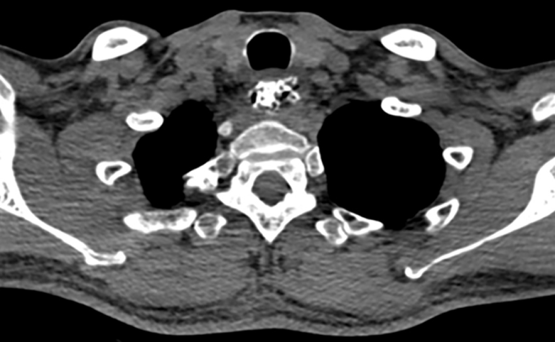
Computed tomography revealed a high-density shadow occupying the entire esophagus, from the seventh cervical vertebra to the second thoracic vertebra, without perforation (Figure 1).
Emergent endoscopy revealed an irregular and sharp-pointed chicken bone tightly embedded in the esophageal wall, with minor bleeding (Figure 2A).
Esophageal injury caused by accidental swallowing and embedding of a chicken bone in the esophageal wall.
The chicken bone was pushed into the stomach cavity and a condom was used to wrap it (Figure 2B). The chicken bone was easily extracted using foreign forceps (Figure 2C).
The patient was discharged the following day with only primary esophageal injuries caused by the chicken bone, and no additional significant damage was found.
Foreign bodies in the digestive tract refer to food or other objects that cannot be digested and are not easily excreted in the patient's digestive tract. In severe cases, it can lead to serious complications such as bleeding and perforation, and even endanger life[4]. Leinwand et al[5] reported 13 cases of the most significant and severe esophageal button battery ingestions that occurred at the authors' institution from 2009 to 2015. Liming et al[6] presented a case where a long-existing esophageal foreign body led to bronchial compression and an acquired tracheoesophageal fistula. Endoscopic removal is the most common used treatment for foreign bodies in the digestive tract[7], which is a procedure that uses endoscopes and related accessories to remove foreign bodies. The types of foreign bodies in the gastrointestinal tract are varied, and we often need to tailor our approach and choose the most appropriate method based on the size, shape, material, and actual situation of the foreign body in clinical practice, thereby reducing complications, effectively avoiding surgery, and even saving the patient's life. The chicken bone in this case was sharp. If it hadn't been removed in time, it would have been very likely to cause esophageal perforation or pierce large blood vessels, resulting in serious consequences[8].
In this case, due to the large size and irregular polyhedral and sharp shape of the bone, forcibly dragging it out of the throat area would extremely likely cause severe damage to the throat and the entrance of the esophagus, leading to massive hemorrhage or perforation. So, we attempt to push it into the stomach cavity firstly. During the process, the chicken bone was clamped and the endoscope was advanced slowly and gently. At the same time, air was continuously injected to maintain a better field of view and a more expanded esophageal cavity to reduce mucosa damage.
Considering that traditional protective devices are not suitable for covering such large and irregular sharp chicken bone, we came up with the idea of using a condom as an alternative. After carefully pushing the bone into the stomach cavity, we use the foreign body forceps to clamp part of the bone and send it into the opening of the condom. Then, clamp the opening edge of the condom and shake it gently so that the whole bone can be completely inside the condom. Using foreign forceps, we easily extracted it by grasping the open edge of the condom, avoiding possible esophageal mucosal injuries, bleeding and perforation.
The patient was discharged the next day with only the primary esophageal injuries caused by the chicken bone and no additional significant damage found, which proves that the method we adopted is successful. Therefore, in cases where traditional protective devices are not suitable for covering large and irregular sharp foreign bodies, a condom can serve as an alternative because of its smooth and oily nature, which offers both mucosal protection and lubrication during the endoscopic removal process.
| 1. | Ikenberry SO, Jue TL, Anderson MA, Appalaneni V, Banerjee S, Ben-Menachem T, Decker GA, Fanelli RD, Fisher LR, Fukami N, Harrison ME, Jain R, Khan KM, Krinsky ML, Maple JT, Sharaf R, Strohmeyer L, Dominitz JA; ASGE Standards of Practice Committee. Management of ingested foreign bodies and food impactions. Gastrointest Endosc. 2011;73:1085-1091. [RCA] [PubMed] [DOI] [Full Text] [Cited by in Crossref: 468] [Cited by in RCA: 498] [Article Influence: 35.6] [Reference Citation Analysis (1)] |
| 2. | Ahmed Z, Arif SF, Ong SL, Badal J, Lee-Smith W, Renno A, Alastal Y, Nawras A, Aziz M. Cap-Assisted Endoscopic Esophageal Foreign Body Removal Is Safe and Efficacious Compared to Conventional Methods. Dig Dis Sci. 2023;68:1411-1425. [RCA] [PubMed] [DOI] [Full Text] [Cited by in Crossref: 3] [Cited by in RCA: 4] [Article Influence: 2.0] [Reference Citation Analysis (0)] |
| 3. | Birk M, Bauerfeind P, Deprez PH, Häfner M, Hartmann D, Hassan C, Hucl T, Lesur G, Aabakken L, Meining A. Removal of foreign bodies in the upper gastrointestinal tract in adults: European Society of Gastrointestinal Endoscopy (ESGE) Clinical Guideline. Endoscopy. 2016;48:489-496. [RCA] [PubMed] [DOI] [Full Text] [Cited by in Crossref: 274] [Cited by in RCA: 384] [Article Influence: 42.7] [Reference Citation Analysis (0)] |
| 4. | Boo SJ, Kim HU. [Esophageal Foreign Body: Treatment and Complications]. Korean J Gastroenterol. 2018;72:1-5. [RCA] [PubMed] [DOI] [Full Text] [Cited by in Crossref: 8] [Cited by in RCA: 9] [Article Influence: 1.3] [Reference Citation Analysis (0)] |
| 5. | Leinwand K, Brumbaugh DE, Kramer RE. Button Battery Ingestion in Children: A Paradigm for Management of Severe Pediatric Foreign Body Ingestions. Gastrointest Endosc Clin N Am. 2016;26:99-118. [RCA] [PubMed] [DOI] [Full Text] [Cited by in Crossref: 100] [Cited by in RCA: 93] [Article Influence: 10.3] [Reference Citation Analysis (1)] |
| 6. | Liming BJ, Fischer A, Pitcher G. Bronchial Compression and Tracheosophageal Fistula Secondary to Prolonged Esophageal Foreign Body. Ann Otol Rhinol Laryngol. 2016;125:1030-1033. [RCA] [PubMed] [DOI] [Full Text] [Cited by in Crossref: 5] [Cited by in RCA: 5] [Article Influence: 0.6] [Reference Citation Analysis (0)] |
| 7. | Sugawa C, Ono H, Taleb M, Lucas CE. Endoscopic management of foreign bodies in the upper gastrointestinal tract: A review. World J Gastrointest Endosc. 2014;6:475-481. [RCA] [PubMed] [DOI] [Full Text] [Full Text (PDF)] [Cited by in CrossRef: 115] [Cited by in RCA: 138] [Article Influence: 12.5] [Reference Citation Analysis (0)] |


