Published online Jan 16, 2025. doi: 10.4253/wjge.v17.i1.102185
Revised: November 27, 2024
Accepted: December 16, 2024
Published online: January 16, 2025
Processing time: 97 Days and 2.9 Hours
Gastric bezoars are indigestible masses that can lead to gastrointestinal ob
A 57-year-old male with a history of type 2 diabetes presented with severe epigastric pain and vomiting. Endoscopy revealed two large phytobezoars and a gastric ulcer. Initial attempts at mechanical fragmentation with a polypectomy snare and Coca-Cola ingestion for dissolution were unsuccessful due to the large size and complex structure of the bezoars. An innovative approach using snare-tip electrocautery was then employed. It successfully penetrated the slippery, hard surface of the bezoars and fragmented them into smaller pieces. The patient was subsequently treated with Coca-Cola ingestion, enzyme supplements, and proton pump inhibitors. He was discharged without complications following the endoscopic sessions.
Snare-tip electrocautery is a safe, cost-effective, and minimally invasive alter
Core Tip: Snare-tip electrocautery provided precise and efficient fragmentation of refractory bezoars, which reduced the need for surgery. Snare-tip electrocautery may be particularly useful in cases unresponsive to conventional treatments.
- Citation: Lim CH, Lim CJ, Yao CT, Chang CC. Novel approach to managing two enormous bezoars with successive snare-tip electrocautery: A case report. World J Gastrointest Endosc 2025; 17(1): 102185
- URL: https://www.wjgnet.com/1948-5190/full/v17/i1/102185.htm
- DOI: https://dx.doi.org/10.4253/wjge.v17.i1.102185
Bezoars are foreign bodies formed within the gastrointestinal tract due to the aggregation of undigested material. They are categorized into four distinct types based on their composition: Phytobezoars; trichobezoars; pharmacobezoars; and lactobezoars[1]. Phytobezoars are prevalent in the stomach. Bezoars exceeding 3 cm in size pose an elevated risk of gastric ulcers and intestinal obstruction. Ulcers are often localized to the angulus of the stomach due to friction from the bezoar. Bezoars are treated with endoscopic mechanical fragmentation and the use of carbonated beverages such as Coca-Cola. Large bezoars typically require combination therapy consisting of multiple treatments over an extended period. The outcomes can be unsatisfactory, especially in cases of symptomatic gigantic bezoars, which often require surgical intervention. Herein, we present the case of two massive stomach bezoars complicated by symptoms of gastric outlet obstruction that were successfully managed using unconventional snare tip electrocautery.
A 57-year-old Asian male presented to our emergency department with severe epigastric pain.
The patient had a medical history of type 2 diabetes. He reported experiencing postprandial epigastric discomfort persisting for over 3 months. Recently, the patient had also began experiencing episodes of postprandial vomiting, even with the ingestion of water.
The patient’s past medical history is unremarkable, with no significant illnesses, hospitalizations, or surgical procedures reported.
Family history of Hypertension in his father.
Physical examination revealed abdominal distension with tenderness localized to the epigastric region.
Laboratory analysis indicated the presence of microcytic anemia, as evidenced by hemoglobin at 9.4 g/dL (normal range: 13-18 g/dL) and mean corpuscular volume of 72.7 fl (normal range: 85.6-102.5 fl).
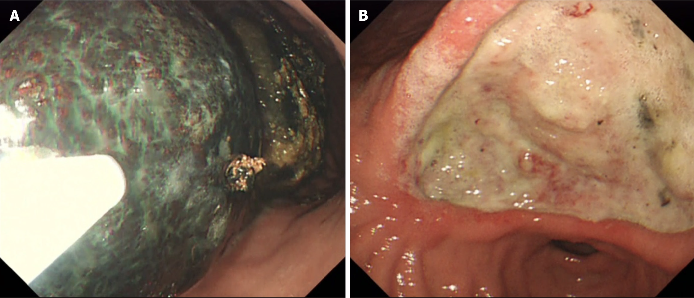
Kidney, ureter, bladder X-ray revealed gastric distension with fecal-like material present. Esophagogastroduodenoscopy revealed the presence of two large phytobezoars, each measuring 8 cm in diameter. They were situated within the fundus and body of the stomach. A sizable deep ulcer (Forrest IIb, 5-6 cm) located in the angular incisure extending into the antrum area was also detected (Figure 1A and B).
Phytobezoar and ulcer.
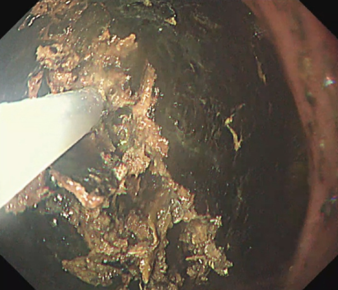
Initial attempts at mechanical lithotripsy utilizing a 33-mm round snare (extra-large rounded, single-use polypectomy snare; Boston Scientific, Marlborough, MA, United States) and raptor forceps were unsuccessful due to the slippery structural density of the bezoar, which prevented effective anchoring of the snare. Subsequent interventions employed the snare tip for electrocautery [forced coagulation mode, effect 2, 35-50 W (VIO 300D; Erbe, Marietta, GA, United States)] extended by approximately 2-4 mm to enable precise bezoar fragmentation while minimizing mucosal contact (Figure 2). The bezoar was reduced to fragments smaller than 2 cm to mitigate the risk of small intestine obstruction (Video).
During hospitalization, the patient’s intake consisted solely of diet Coca-Cola (600-1000 mL every 6 hours) and a digestive enzyme supplement. Proton pump inhibitor therapy was initiated to address ulceration. Eight days after the initial treatment, a follow-up esophagogastroduodenoscopy demonstrated incomplete dissolution of the bezoar. There was evidence of the original unfragmented mass and the aggregation of fragmented remnants into a smaller concretion.
Subsequent endoscopic sessions addressed the remaining unfragmented bezoar. The day following the final endoscopic fragmentation, the patient was discharged uneventfully.
Coca-Cola is the primary treatment for the dissolution of gastric bezoars[2]. Alternatively, combined endoscopic mechanical fragmentation using lithotripters, snares, or forceps may be employed. Giant bezoars refractory to conventional treatments often require surgical intervention. Argon plasma coagulation (APC) and electrohydraulic lithotripsy have also been used to manage massive bezoars. However, only limited case series data and specific procedural reports are available[3,4].
During APC or laser lithotripsy treatment, accurately assessing the depth of fragmentation can be challenging, and the soft nature of the APC probe complicates effective manipulation of the bezoar. Similarly, electrohydraulic lithotripsy introduces a significant amount of water into the gastric lumen, which can obscure the visualization of the bezoar and surrounding mucosa, increasing the risk of gastric mucosal injury. Moreover, these methods often require additional tools, such as snares or forceps, to fragment debris effectively. Both techniques not only fail to fragment bezoars independently and are time-consuming but also pose an increased risk of mucosal injury due to limited visualization of treatment depth.
In this case, snare-tip electrocautery offers a viable alternative for managing intractable bezoars, particularly in resource-limited settings where advanced equipment may not be available. Extending the snare tip approximately 0.2-0.4 cm beyond the snare sheath, similar to techniques used with a longer needle knife, allows for precise visualization of the cutting depth and helps prevent mucosal injury. Additionally, the snare sheath facilitates repositioning of the bezoar in challenging angles, enhancing procedural effectiveness. The electrocautery function reduces the stickiness of clay-like bezoars, which can otherwise impede fragmentation, enabling more efficient treatment. The same snare can also be used to fragment residual bezoar debris effectively.
Snare-tip electrocautery is an effective, safe, and cost-efficient non-surgical intervention for large, refractory bezoars. Its versatility enables precise depth control in confined spaces, and the procedure time is reduced. Snare-tip electrocautery is a valuable endoscopic option for managing refractory bezoars.
| 1. | Iwamuro M, Okada H, Matsueda K, Inaba T, Kusumoto C, Imagawa A, Yamamoto K. Review of the diagnosis and management of gastrointestinal bezoars. World J Gastrointest Endosc. 2015;7:336-345. [RCA] [PubMed] [DOI] [Full Text] [Full Text (PDF)] [Cited by in CrossRef: 214] [Cited by in RCA: 199] [Article Influence: 19.9] [Reference Citation Analysis (5)] |
| 2. | Liu FG, Meng DF, Shen X, Meng D, Liu Y, Zhang LY. Coca-Cola consumption vs fragmentation in the management of patients with phytobezoars: A prospective randomized controlled trial. World J Gastrointest Endosc. 2024;16:83-90. [RCA] [PubMed] [DOI] [Full Text] [Full Text (PDF)] [Cited by in RCA: 3] [Reference Citation Analysis (0)] |
| 3. | Curcio G, Granata A, Azzopardi N, Barresi L, Tarantino I, Traina M. Argon plasma coagulation before mechanical fragmentation of a large gastric persimmon bezoar: the woodworm technique. Endoscopy. 2013;45:E227-E228. [RCA] [PubMed] [DOI] [Full Text] [Cited by in Crossref: 1] [Cited by in RCA: 1] [Article Influence: 0.1] [Reference Citation Analysis (0)] |
| 4. | Jinushi R, Yano T, Imamura N, Ishii N. Endoscopic treatment for a giant gastric bezoar: Sequential use of electrohydraulic lithotripsy, alligator forceps, and snares. JGH Open. 2021;5:522-524. [RCA] [PubMed] [DOI] [Full Text] [Full Text (PDF)] [Cited by in Crossref: 3] [Cited by in RCA: 3] [Article Influence: 0.8] [Reference Citation Analysis (0)] |


