Copyright
©The Author(s) 2023.
World J Gastrointest Endosc. Nov 16, 2023; 15(11): 666-675
Published online Nov 16, 2023. doi: 10.4253/wjge.v15.i11.666
Published online Nov 16, 2023. doi: 10.4253/wjge.v15.i11.666
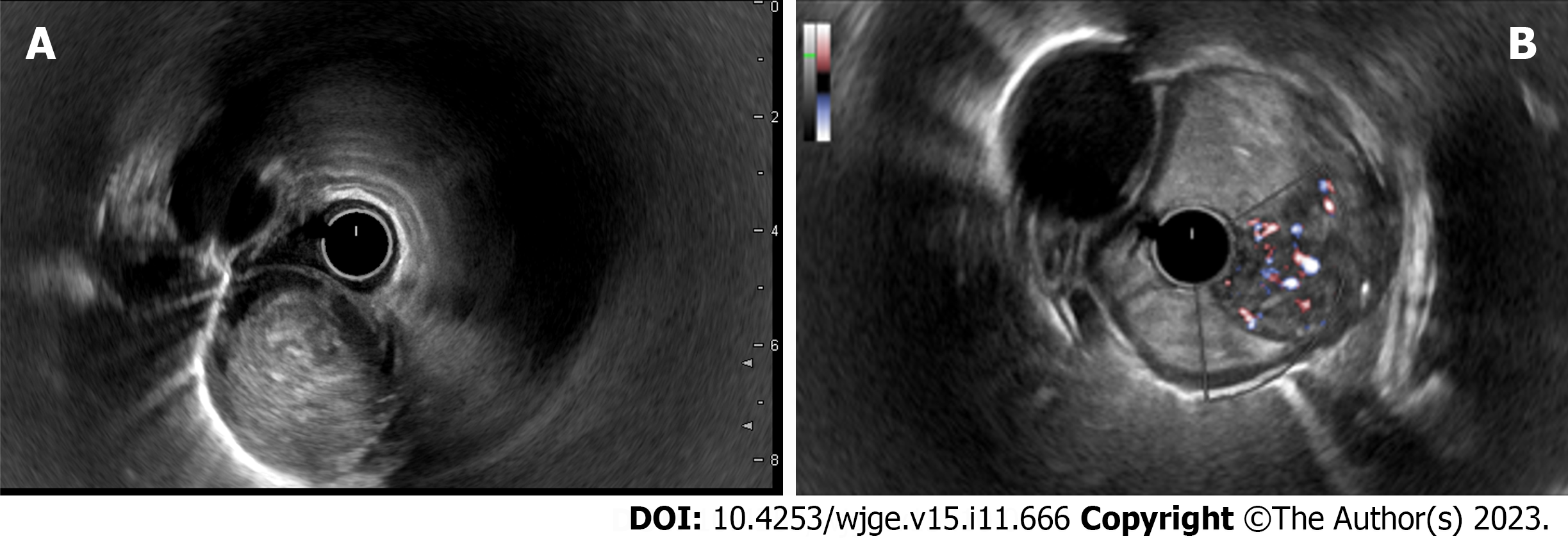
Figure 2 Endosonography of the esophagus.
A: A heterogeneous hypoechoic mass with a smooth and clear borders, cylindrically shaped, originating from submucosal layer (3rd echo layer); B: A doppler color mode shows a hypervascular zone at the base of the tumor with multiple large feeding vessels, up to 4-5 mm in diameter, extending along the wall of the esophagus.
- Citation: Dzhantukhanova S, Avetisyan LG, Badakhova A, Starkov Y, Glotov A. Hybrid laparo-endoscopic access: New approach to surgical treatment for giant fibrovascular polyp of esophagus: A case report and review of literature. World J Gastrointest Endosc 2023; 15(11): 666-675
- URL: https://www.wjgnet.com/1948-5190/full/v15/i11/666.htm
- DOI: https://dx.doi.org/10.4253/wjge.v15.i11.666


