Copyright
©The Author(s) 2018.
World J Gastrointest Endosc. Oct 16, 2018; 10(10): 283-293
Published online Oct 16, 2018. doi: 10.4253/wjge.v10.i10.283
Published online Oct 16, 2018. doi: 10.4253/wjge.v10.i10.283
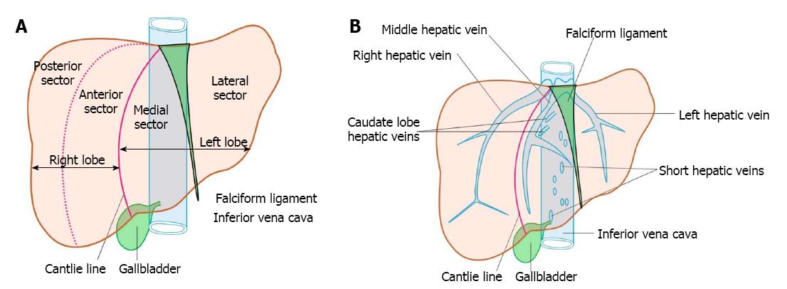
Figure 1 Anatomy of liver.
A: The four sectors of liver, i.e., right anterior, right posterior, left medial and left lateral; B: The three major veins, emerge from the posterior surface of the liver and open immediately into the supra hepatic part of inferior vena cava (IVC) just before it pierces the diaphragm. Short hepatic/accessory veins drain into lower part of IVC. The accessory veins and the caudate lobe veins join the anterior and lateral aspect of IVC.
- Citation: Sharma M, Somani P, Rameshbabu CS. Linear endoscopic ultrasound evaluation of hepatic veins. World J Gastrointest Endosc 2018; 10(10): 283-293
- URL: https://www.wjgnet.com/1948-5190/full/v10/i10/283.htm
- DOI: https://dx.doi.org/10.4253/wjge.v10.i10.283


