Copyright
©The Author(s) 2017.
World J Gastrointest Endosc. Aug 16, 2017; 9(8): 417-424
Published online Aug 16, 2017. doi: 10.4253/wjge.v9.i8.417
Published online Aug 16, 2017. doi: 10.4253/wjge.v9.i8.417
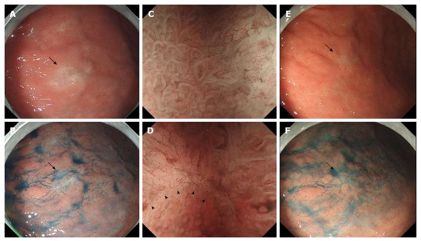
Figure 1 Endoscopic imaging of the antral lesion before and after the eradication.
A: An endoscopic image before the eradication shows a 13-mm whitish/slightly brownish lesion (arrow) on the lesser curvature of the antrum; B: Indigo carmine dye spray reveals that the antral lesion (arrow) is almost flat; C: Magnification endoscopy with narrow-band imaging shows the lesion (C, right side) exhibits loss or irregularity of the microsurface pattern and irregular microvascular proliferation compared to the background mucosa (C, left side). A demarcation line (D, arrowheads) is seen at the periphery of the lesion. E-F: Endoscopic images after the eradication therapy exhibit that the mucosal lesion (arrow) decreases in size to 7 mm in diameter (E) and preserves the gross appearance with indigo carmine dye spray (F).
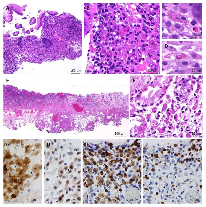
Figure 2 Pathological features of the antral lesion.
The biopsy specimen stained by hematoxylin and eosin shows chronic gastritis with crowded granulated cells (A, B). The histiocytoid cells have fine eosinophilic granules similar to those of eosinophils (C). Intranuclear cytoplasmic granules suggesting Dutcher bodies are observed (D). E-F: The ESD specimen stained by hematoxylin and eosin shows that the lesion (E, indicated by a dotted line) is edematous and demarcated and includes many granulated cells (F). G-J: Immunohistochemical (G-H) and in situ hybridization (I-J) findings with ESD specimens show that the granulated cells (arrowheads) are positive for CD79a (G), multiple myeloma oncogene 1 (H), and kappa (I) and negative for lambda (J), while plasma cells without cytoplasmic granules are polyphenotypic.
- Citation: Yorita K, Iwasaki T, Uchita K, Kuroda N, Kojima K, Iwamura S, Tsutsumi Y, Ohno A, Kataoka H. Russell body gastritis with Dutcher bodies evaluated using magnification endoscopy. World J Gastrointest Endosc 2017; 9(8): 417-424
- URL: https://www.wjgnet.com/1948-5190/full/v9/i8/417.htm
- DOI: https://dx.doi.org/10.4253/wjge.v9.i8.417


