Copyright
©The Author(s) 2017.
World J Gastrointest Endosc. May 16, 2017; 9(5): 220-227
Published online May 16, 2017. doi: 10.4253/wjge.v9.i5.220
Published online May 16, 2017. doi: 10.4253/wjge.v9.i5.220
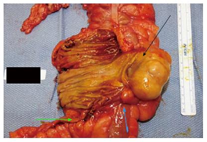
Figure 1 Cecal mass (black arrow, lead point) pulling the terminal ileum (blue arrow) causing intermittent intussusception, appendix is depicted by yellow arrow.
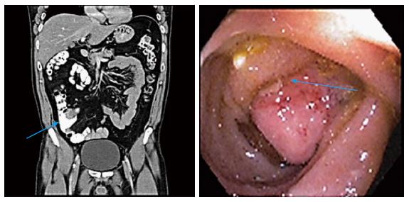
Figure 2 Terminal ileal mass intussusception into the cecum.
Computed tomography (CT) scan shows the mass, colonoscopy view confirms the CT finding. Both depicted with a blue arrow.
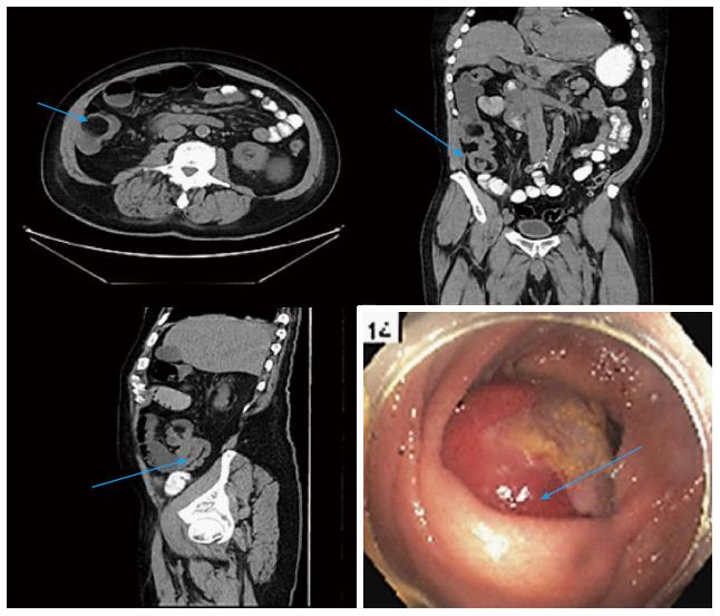
Figure 3 Ileocecal Intussusception.
Computed tomography scans (axial, sagittal, coronal) shows the terminal ileum intussusception in the cecum. Colonoscopy confirms the mass protruding into the cecum.
Figure 4 Ileocecal intussusception.
Computed tomography scans (axial, sagittal, coronal) shows the mass intussusception in the cecum. Colonoscopy confirms the mass protruding through the ileocecal valve.
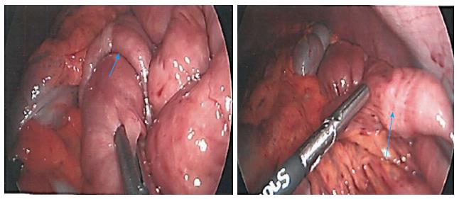
Figure 5 Laparoscopic view of jejunal-jejunal intussusception which was reduced.
Physiologic peristalsis, idiopathic finding.
- Citation: Shenoy S. Adult intussusception: A case series and review. World J Gastrointest Endosc 2017; 9(5): 220-227
- URL: https://www.wjgnet.com/1948-5190/full/v9/i5/220.htm
- DOI: https://dx.doi.org/10.4253/wjge.v9.i5.220


