Copyright
©The Author(s) 2017.
World J Gastrointest Endosc. Mar 16, 2017; 9(3): 127-132
Published online Mar 16, 2017. doi: 10.4253/wjge.v9.i3.127
Published online Mar 16, 2017. doi: 10.4253/wjge.v9.i3.127
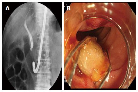
Figure 1 Endoscopic retrograde cholangiopancreatography on bile duct stone in athe patient with Billroth II-reconstructed stomach.
A: Stones, sized 7 mm, were observed by cholangiography; B: Stones were gripped with a basket, and collection of stone was successful.
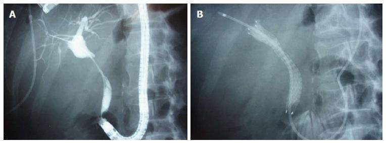
Figure 2 Endoscopic retrograde cholangiopancreatography on obstructive jaundice in the a patient with Billroth II-reconstructed stomach.
A: Intense stenosis was observed from the upper through middle bile duct by cholangiography; B: The endoscopic nasobiliary drainage tube was placed after placement of a metallic stent.
- Citation: Sakai Y, Tsuyuguchi T, Mikata R, Sugiyama H, Yasui S, Miyazaki M, Yokosuka O. Utility of endoscopic retrograde cholangiopancreatography on biliopancreatic diseases in patients with Billroth II-reconstructed stomach. World J Gastrointest Endosc 2017; 9(3): 127-132
- URL: https://www.wjgnet.com/1948-5190/full/v9/i3/127.htm
- DOI: https://dx.doi.org/10.4253/wjge.v9.i3.127


