Copyright
©The Author(s) 2016.
World J Gastrointest Endosc. Nov 16, 2016; 8(19): 690-696
Published online Nov 16, 2016. doi: 10.4253/wjge.v8.i19.690
Published online Nov 16, 2016. doi: 10.4253/wjge.v8.i19.690
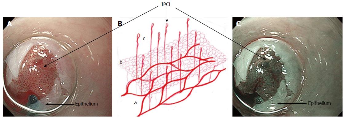
Figure 1 Mucosal vessels.
A and C: Endoscopic images during per-oral endoscopic myotomy procedure (high magnification images); after unintentional removal of the epithelium (white layer), top half of epithelium was peeled off, and IPCLs were exposed. IPCLs appear as regularly-arranged, red dots (A: White light) or dark green spots (C: NBI); B: A schematic representation of the vascular network of esophageal mucosa: a: Branching vessels; b: SECN; c: IPCL. IPCL: Intrapapillary capillary loop; SECN: Sub epithelial capillary network; NBI: Narrow band imaging.
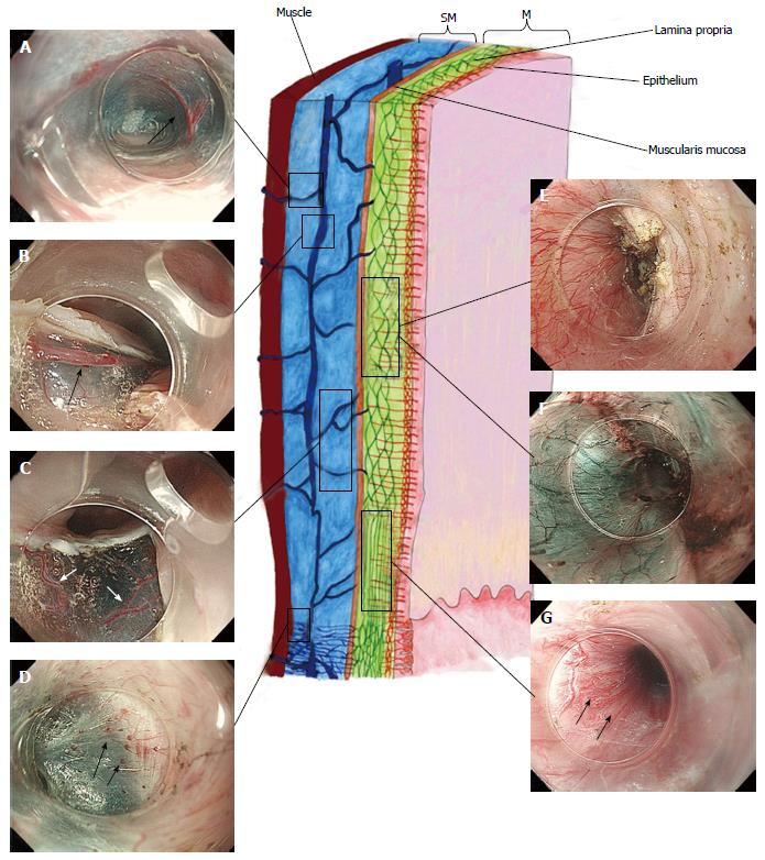
Figure 2 Esophageal wall and esophago-gastric junction vasculature: Schematic illustration and endoscopic corresponding images (high magnification images).
Black arrow indicates vessels. This image was originally published in “Treatment Strategies Gastroenterology”[28]. A: Perforating vessels from the outer esophagus to the submucosal vessel; image captured during tunnelization in POEM (bottom side muscle layer, left side submucosal lifting); B: Submucosal drainage vessel (mucosal layer lifted on during ESD). These veins can become esophageal varices in portal hypertension; C: Submucosal vessels connecting the drainage veins to the mucosal branching vessels (in the lamina propria); D: Spindle veins immediately below the GEJ (in left side of the image, in blue, the submucosa and in the right side the muscle); E and F: Whitet light and NBI of the branching vessels (seen from inside the submucosal tunnel). Backside of the mucosa on the left; muscle-already cut-on the right; G: Passage between lower esophagus and GEJ. In the image is possible to recognize, in different planes, all the vessel of the submucosa and lamina propria (palisade vessels). POEM: Per-oral endoscopic myotomy; ESD: Endoscopic submucosal dissection; GEJ: Esophagogastric junction; NBI: Narrow band imaging; M: Mucosa; SM: Submucosa.
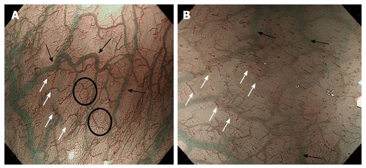
Figure 3 High magnifying narrow band imaging image of normal esophageal mucosa (luminal side).
A: Soft pressure of the endoscope distal attachment (“hood”) onto the mucosal surface demonstrates SECN, hard pressure onto the mucosa compresses horizontal vessels, allowing clear observation of IPCLs; B: In the circle the SECN located at the top layer of lamina propria mucosae, just beneath the epithelium. The black arrows indicate the branching vessels into the lower lamina propria; white arrows indicate the IPCL located in the epithelial papilla, which is a projection of lamina propria mucosae into the epithelium. SECN: Sub-epithelial capillary network; IPCL: Intrapapillary capillary loop.
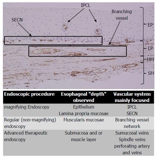
Figure 4 The figure shows the histology of a non-pathologic esophageal specimen.
The vessels’ wall has been colored by CD34, showing superficially the IPCLs (upper part of the lamina propria, arising the epithelium) and the SECN; deeply in the lamina propria the branching vessels. In the sumucosal layer also the drainage veins are evident. The table summarizes the vascular system observed and its own esophageal layer according to the different endoscopic procedure performed. SECN: Sub-epithelial capillary network; IPCL: Intrapapillary capillary loop; EP: Epithelium; LP: Lamina propria; MM: Muscolaris mucosa; SM: Submucosa.
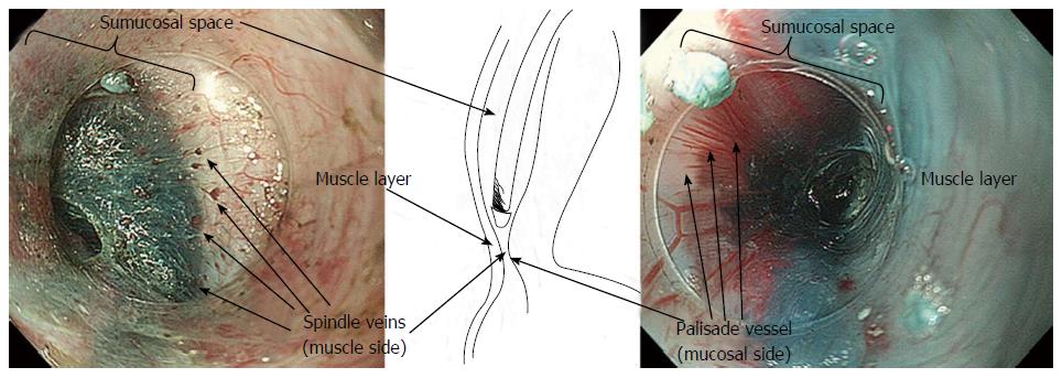
Figure 5 In the center a scheme of the submucosal view at the gastro-esophageal junction during per-oral endoscopic myotomy.
At the muscle side (left endoscopic image) the spindle vein are clearly visible; at the mucosal side (seen on its backside, right endoscopic image) the palisade vessel are recognized. High magnification images.
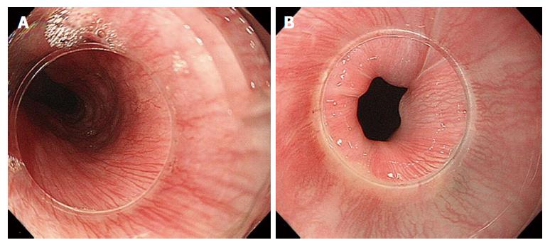
Figure 6 Palisade vessels at the esophageal sphincter.
A: Palisade vessels at the upper esophageal sphincter; B: In the lower esophageal sphincter, the vessels, located in the lamina propria, are continuation of the branching vessels, “stretched” by the high pressure present in the area. High magnification images.
- Citation: Maselli R, Inoue H, Ikeda H, Onimaru M, Yoshida A, Santi EG, Sato H, Hayee B, Kudo SE. Microvasculature of the esophagus and gastroesophageal junction: Lesson learned from submucosal endoscopy. World J Gastrointest Endosc 2016; 8(19): 690-696
- URL: https://www.wjgnet.com/1948-5190/full/v8/i19/690.htm
- DOI: https://dx.doi.org/10.4253/wjge.v8.i19.690


