Copyright
©The Author(s) 2016.
World J Gastrointest Endosc. May 25, 2016; 8(10): 402-408
Published online May 25, 2016. doi: 10.4253/wjge.v8.i10.402
Published online May 25, 2016. doi: 10.4253/wjge.v8.i10.402
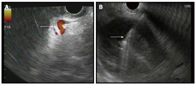
Figure 1 Endoscopic ultrasound of walled off necrosis.
A: Doppler used to visualize any interventing vessels (arrow) including varices; B: FNA needle (arrow) seen entering necrotic cyst under EUS guidance. EUS: External urethral sphincter; FNA: Fine-needle aspiration.
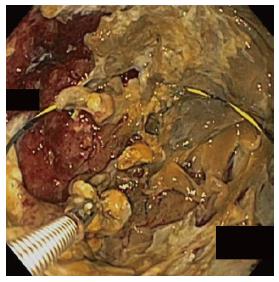
Figure 2 Endoscopic necrosectomy performed with debridement of the cyst cavity.
Wire is seen coiled within the cyst to maintain access through the procedure.
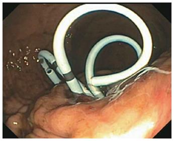
Figure 3 Pigtail stents left in place at the end of endoscopic necrosectomy to encourage ongoing drainage.
Figure 4 Varices identified through various methods.
A: Large gastric varix (arrow) seen endoscopically; B: Peri-gastric varix (arrow) seen within the cyst cavity during endoscopic necrosectomy; C: Computed tomography scan showing gastric varices (arrows) in close proximity to the stomach (S) and walled off necrosis (WON).
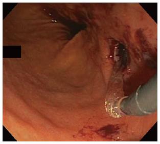
Figure 5 Status-post balloon dilation of the necrosectomy tract, shown with self-limited bleeding.
- Citation: Storm AC, Thompson CC. Safety of direct endoscopic necrosectomy in patients with gastric varices. World J Gastrointest Endosc 2016; 8(10): 402-408
- URL: https://www.wjgnet.com/1948-5190/full/v8/i10/402.htm
- DOI: https://dx.doi.org/10.4253/wjge.v8.i10.402


