Copyright
©The Author(s) 2015.
World J Gastrointest Endosc. Jul 25, 2015; 7(9): 860-871
Published online Jul 25, 2015. doi: 10.4253/wjge.v7.i9.860
Published online Jul 25, 2015. doi: 10.4253/wjge.v7.i9.860
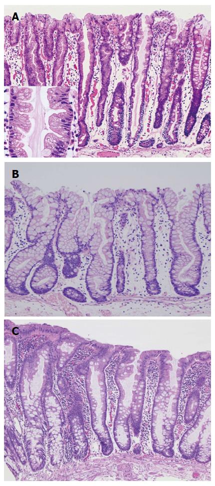
Figure 1 Histological findings of hyperplastic polyps.
A: Microvesicular hyperplastic polyp (MVHP): The crypts and surface epithelium showing a serrated appearance with micro-goblet cells increased. High power view is shown at left side bottom. Many small droplet (microvesicular) mucin within the cytoplasm at the epithelial layer is specific findings as shown the picture; B: Goblet-cell rich HP: In contrast to MVHP, this type polyp is showing a much less serrated appearance inside the surface epithelium of crypts. And showing a preponderance of goblet cells without microvesicular mucin; C: Mucin-poor HP (MPHP): MPHP is rare, and little is known about their molecular features and natural history. The histological features are showing no cytoplasmic mucin with a luminal serration pattern. And also showing increased nuclear atypia without pseudostratification.
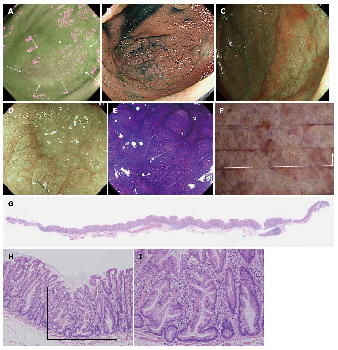
Figure 2 A case of sessile serrated adenoma/polyp without cytological dysplasia (scope: CF: FH260AZI).
A: AFI imaging. The flat elevated polyp is approximately 37 mm in diameter as is located in cecum. No change to magenta of the tumor relative to the surrounding normal mucosa can be observed (inside white arrows); B: Indigocarmine spraying endoscopic finding. The structure of the granular surface is clearly revealed by chromoendoscopy; C: NBI observation, non-magnified. A red cap is covering the surface of the tumor; D: NBI observation, magnified. Small black dots can be observed in the tumor. This finding indicates that this tumor possesses the characteristic of SSA/P; E: Crystal violet staining under magnified observation. Type II open pits (II-O pits) containing with normal type II pits are shown in the tumor; F: Stereoscopic finding. The tumor was excised by the ESD method. The tumor was cut into12 pieces; G: HE staining, whole specimen findings from section #4; H: Low power view of the HE staining findings. The tumor contains serrated glands in the mucosal layer; I: High power view of the HE staining findings. Typical histological findings for SSA/P. The crypt exhibits an “inverted T” type. NBI: Narrow band imaging; SSA/P: Sessile serrated adenoma/polyp; AFI: Auto fluorescence imaging.
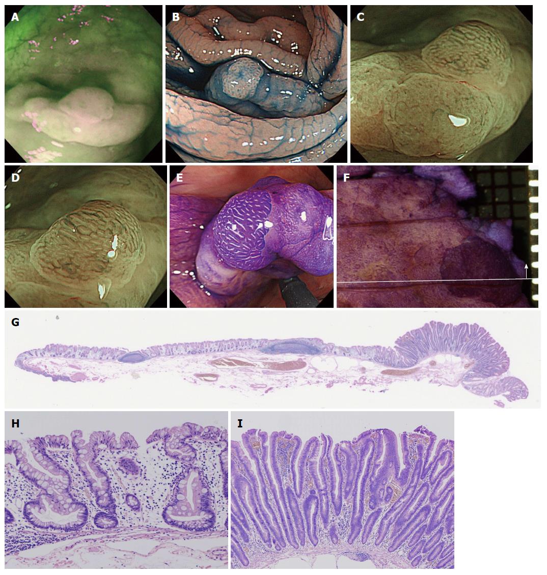
Figure 3 A case of sessile serrated adenoma/polyp with cytological dysplasia (scope: CF: FH260AZI).
A: AFI imaging. The polyp is shown as a flat elevated lesion with a small nodule and is located in the ascending colon. A slightly change to a magenta color can be seen localized to a small elevated lesion in the tumor; B: Indigocarmine spraying endoscopic finding. The small elevated nodule in the tumor can be seen observed following dye spraying; C: Magnified NBI observation. In the tumor lesion, whitish mucosa with II-D pits can be observed. The microcapillary vessels are not dilated in the tumor; D: Magnified NBI observation. In contrast, the microcapillary vessels are dilated surrounding the tumor pits at the small elevated nodule. Moreover, a IIIL pit (white line) can be indirectly observed; E: Magnified crystal violet staining observation. Type II open pits (II-O pits) containing normal type II pits are shown in the tumor; F: Stereoscopic finding. The tumor was excised by the ESD method. The tumor was cut eight pieces; G: HE staining, whole specimen findings from section #4 including a small nodule; H: High power view of the HE staining finding. A part of an SSA/P is shown in the picture; I: High power view of the HE staining finding. The small elevated lesion is shown as a neoplastic change. Low grade cytologic dysplasia is present with nuclear hyperchromasia and pseudostratification. NBI: Narrow band imaging; SSA/P: Sessile serrated adenoma/polyp; AFI: Auto fluorescence imaging.
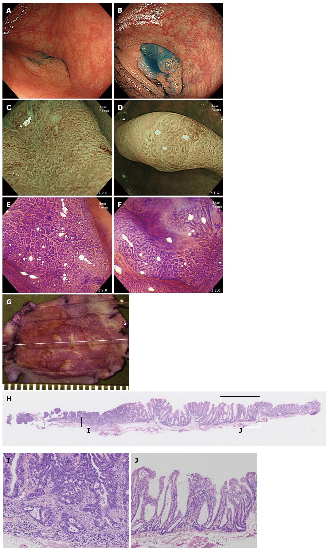
Figure 4 A case of an sessile serrated adenoma/polyp that has invaded the submucosal layer (scope: CF: HQ290I).
A: Conventional white light observation. A flat elevated polyp of approximately 20 mm with a reddish depressed area can be observed in the ascending colon; B: Indigocarmine spraying endoscopic finding. Chromoendoscopy revealed this lesion, which is clearly composed of lesions. One edge area is covered with thick mucus; C: Magnified NBI observation. Firmly attached mucus can be observed on the tumor. A II-D pit that is indicative are markedly dilated crypts can be seen in this area; D: Magnified NBI observation. A granular surface pattern with dilated microcapillary vessels can be observed on this tumor in the absence of a thick mucous adhesion; E and F: Magnified crystal violet staining observation; G: Stereoscopic finding. The tumor was excised by the EMR method. The tumor was cut into seven pieces; H: HE staining, whole specimen finding from #4; I: High power view of the HE staining. The neoplastic glands have invaded into the SM layer to a depth of approximately 400 μm. The glands exhibit high grade dysplastic change; J: Low power view of the HE staining. This polyp is composed of SSA/P glands with markedly dilated crypts. NBI: Narrow band imaging; SSA/P: Sessile serrated adenoma/polyp.
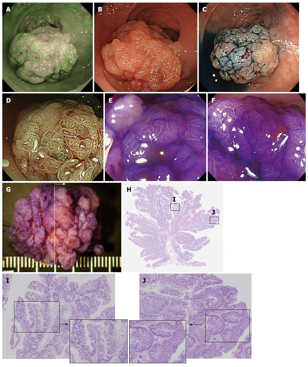
Figure 5 A case of a traditional serrated adenoma with conventional dysplasia (scope: CF: FH260AZI).
A: AFI imaging. A dark green tone that is nearly the same as the surrounding normal colon mucosa can be observed in the tumor; B: Conventional white light observation. A large (approximately 30 mm) semipedunculated polyp exhibiting a slightly reddish change can be observed at the rect-sigmoid junction. There are no findings suggestive of submucosal invasion of the cancer; C: Indigocarmine spraying endoscopic findings. The structure of the nodular surface pattern is clearly revealed; D: NBI observation, magnified. A granular surface pattern with dilated microcapillary vessels can be observed in the tumor; E and F: Magnified crystal violet staining with observation. A type IIIH or IVH pit pattern is shown in the tumor; G: Stereoscopic finding. The tumor was excised by the EMR method. The tumor was cut into 4 pieces; H: HE staining, whole specimen finding from section #2; I: Histological findings from the HE staining. The tumor contains serrated glands in the mucosal layer. Dysplastic change is not observed; J: Histological findings of the HE staining. At several points, TSAs with conventional epithelial dysplasia exhibiting enlarged crowding and pseudostratification of the nuclei with crypt structure dysplastic changes can be observed. TSA: Traditional serrated adenoma; NBI: Narrow band imaging; AFI: Auto fluorescence imaging.
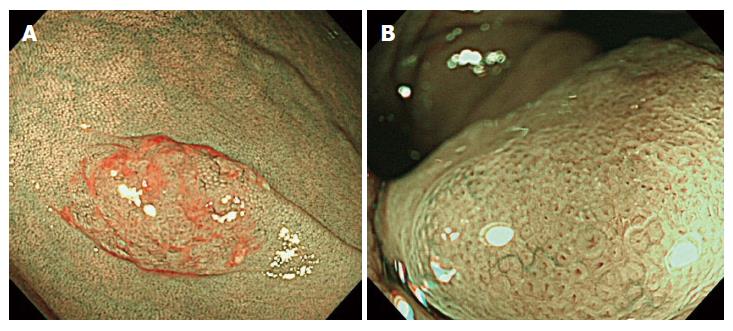
Figure 6 Endoscopic characteristics on narrow band imaging observation.
A: Red cap sign – positive case; B: A finding of showing II-D pit.
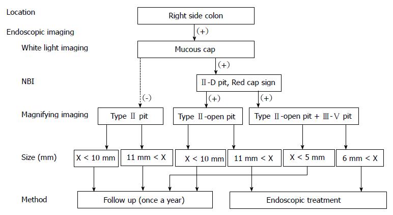
Figure 7 Flow chart for endoscopic treatment about sessile serrated adenoma/polyp.
NBI: Narrow band imaging.
- Citation: Saito S, Tajiri H, Ikegami M. Serrated polyps of the colon and rectum: Endoscopic features including image enhanced endoscopy. World J Gastrointest Endosc 2015; 7(9): 860-871
- URL: https://www.wjgnet.com/1948-5190/full/v7/i9/860.htm
- DOI: https://dx.doi.org/10.4253/wjge.v7.i9.860


