Copyright
©The Author(s) 2015.
World J Gastrointest Endosc. Jul 10, 2015; 7(8): 830-832
Published online Jul 10, 2015. doi: 10.4253/wjge.v7.i8.830
Published online Jul 10, 2015. doi: 10.4253/wjge.v7.i8.830
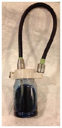
Figure 1 0.
2% indigocarmine solution prepared in the water bottle.
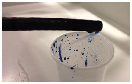
Figure 2 Indigocarmine dye application through the air/water channel of the endoscope.
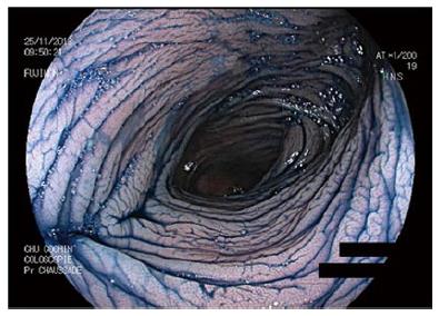
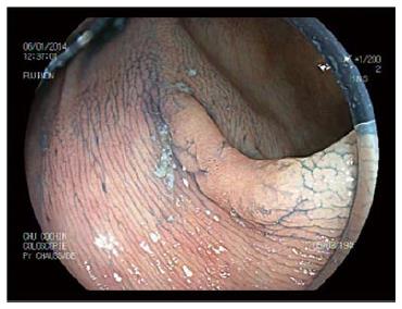
Figure 3 Endoscopic view of the colonic mucosa after indigocarmine staining using a Fujifilm® colonoscope.
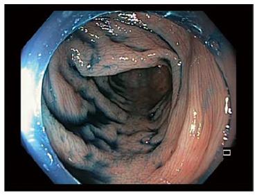
Figure 4 Endoscopic view of the colonic mucosa after indigocarmine staining using an Olympus® colonoscope and a cap.
Figure 5 Endoscopic view of a sessile serrated adenoma after indigocarmine staining.
- Citation: Barret M, Camus M, Leblanc S, Coriat R, Prat F, Chaussade S. Toward an easier indigocarmine chromoendoscopy. World J Gastrointest Endosc 2015; 7(8): 830-832
- URL: https://www.wjgnet.com/1948-5190/full/v7/i8/830.htm
- DOI: https://dx.doi.org/10.4253/wjge.v7.i8.830


