Copyright
©The Author(s) 2015.
World J Gastrointest Endosc. Jul 10, 2015; 7(8): 758-768
Published online Jul 10, 2015. doi: 10.4253/wjge.v7.i8.758
Published online Jul 10, 2015. doi: 10.4253/wjge.v7.i8.758
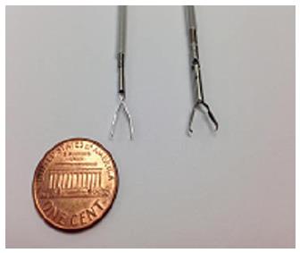
Figure 1 Examples of through the scope clips prior to deployment.
Left: QuickClip 2 (Olympus Medical Systems Co., Tokyo, Japan); Right: Resolution Clip (Boston Scientific, Marlborough, MA).
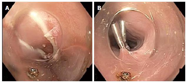
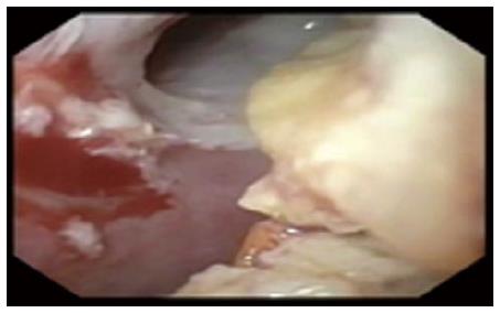
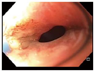
Figure 2 Endoscopic view of mucosotomy during peroral endoscopic myotomy.
A: Esophageal mucosal defect after completion of peroral endoscopic myotomy; B: Defect closed with sequentially placed through the scope clips.
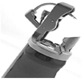
Figure 3 Endoscopic suturing device (Overstitch, Apollo Endosurgery, Austin, TX).
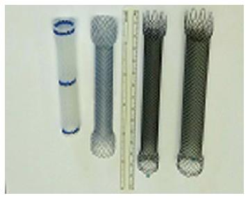
Figure 4 Examples of endoscopic stents.
From Left: Fully covered plastic stent, fully covered metal stent, partially covered metal stent, larger diameter partially covered metal stent.
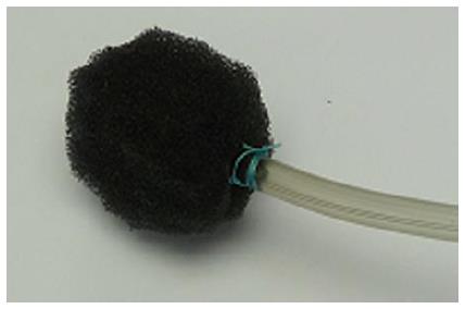
Figure 5 Vacuum-assisted closure device constructed of porous sponge and sutured to a nasogastric feeding tube.
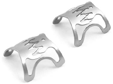
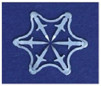
Figure 6 Examples of the over the scope clips (Ovesco Endoscopy, Tubingen, Germany).
Figure 7 Example of the Padlock over the scope clips (Aponos Medical, Kingston, NH).
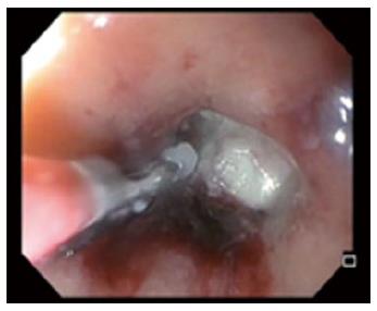
Figure 8 Endoscopic removal of suture foreign body at the opening of a rectal stump fistula.
Figure 9 Undigested food within an esophago-cutaneous fistula tract.
Figure 10 Circumferential argon plasma coagulation catheter ablating epithelialized fistula tract.
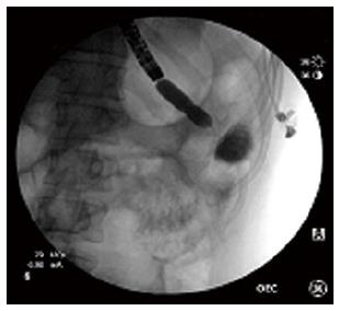
Figure 11 Endoscopic balloon-dilation of a stenotic gastrojejunostomy with adjacent jejunoanastomotic fistula.
- Citation: Winder JS, Pauli EM. Comprehensive management of full-thickness luminal defects: The next frontier of gastrointestinal endoscopy. World J Gastrointest Endosc 2015; 7(8): 758-768
- URL: https://www.wjgnet.com/1948-5190/full/v7/i8/758.htm
- DOI: https://dx.doi.org/10.4253/wjge.v7.i8.758


