Copyright
©The Author(s) 2015.
World J Gastrointest Endosc. Jun 25, 2015; 7(7): 741-746
Published online Jun 25, 2015. doi: 10.4253/wjge.v7.i7.741
Published online Jun 25, 2015. doi: 10.4253/wjge.v7.i7.741
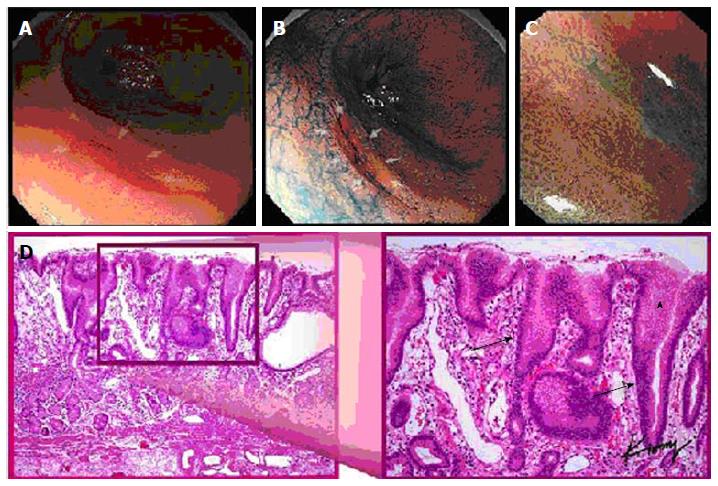
Figure 1 Multiple views of a signet ring cell gastric carcinoma in a single patient: (A) standard white light endoscopy, (B) chromoendoscopy, (C) magnification endoscopy, and (D) histopathology demonstrating elongated gastric glands (arrows) infiltrated with tumor cell (arrowhead).
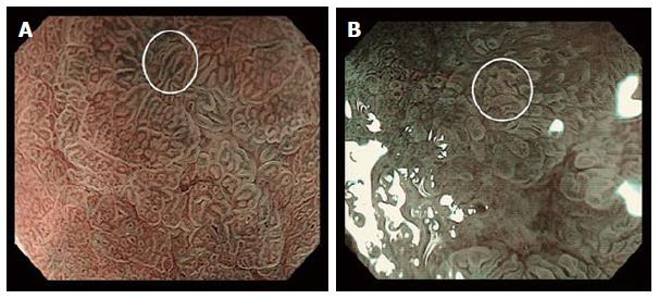
Figure 2 Magnification endoscopy of the stomach: (A) normal polygonal architecture (bottom left, underlying “a”) and a signet ring cell gastric carcinoma demonstrating an elongated or “stretched” gastric gland (white circle); (B) a non-signet ring cell adenocarcinoma demonstrating irregular (non-polygonal) but non-elongated glands (white circle).
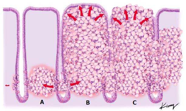
Figure 3 Theoretical view of the pathophysiology of signet ring cell differentiation: (A) tumor cells originating in the neck of the gland and spreading to the submucosal space; (B) an increasing number of tumor cells being packed together, resulting in a barrel shape; and (C) the previously non-exposed tumor becoming exposed through necrosis and formation of an ulcer.
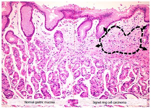
Figure 4 Microscopic view of a signet ring cell gastric carcinoma, demonstrating: (1) normal appearing gastric mucosa (left); and (2) signet ring cells (black dashed circle) causing distortion of the gastric glands (right), consistent with the endoscopic finding of the “stretch sign.
”
- Citation: Phalanusitthepha C, Grimes KL, Ikeda H, Sato H, Sato C, Hokierti C, Inoue H. Endoscopic features of early-stage signet-ring-cell carcinoma of the stomach. World J Gastrointest Endosc 2015; 7(7): 741-746
- URL: https://www.wjgnet.com/1948-5190/full/v7/i7/741.htm
- DOI: https://dx.doi.org/10.4253/wjge.v7.i7.741


