Copyright
©The Author(s) 2015.
World J Gastrointest Endosc. Apr 16, 2015; 7(4): 396-402
Published online Apr 16, 2015. doi: 10.4253/wjge.v7.i4.396
Published online Apr 16, 2015. doi: 10.4253/wjge.v7.i4.396
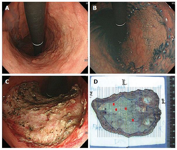
Figure 1 A lesion with a histologic upgraded from extended low-grade dysplasia to adenocarcinoma following endoscopic submucosal dissection.
A: White light endoscopy reveals a large elevated mucosal lesion with nodularity in the lesser curvature side of the body. This lesion was diagnosed as LGD by the endoscopic forceps biopsy; B: This lesion is removed by ESD; C: A large mucosal defect is noted over the gastric body after ESD; D: Mapping of the resected specimen. The tumor size is 75 mm, focal cancer lesions (red bar) mixed with LGD are evident. The lateral and vertical margins are free from tumor. LGD: Low-grade dysplasia; ESD: Endoscopic submucosal dissection.
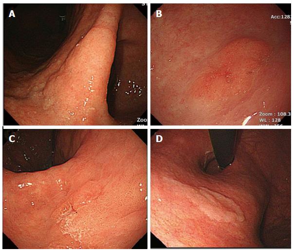
Figure 2 Endoscopic images of biopsy-proven low-grade dysplasia.
A-C: lesion size > 2 cm (A), surface erythema (B), and depressed appearance (C) are endoscopic risk factors for an upgraded histology after endoscopic resection; D: In contrast, the presence of whitish discoloration was a negative factor.
- Citation: Kim JW, Jang JY. Optimal management of biopsy-proven low-grade gastric dysplasia. World J Gastrointest Endosc 2015; 7(4): 396-402
- URL: https://www.wjgnet.com/1948-5190/full/v7/i4/396.htm
- DOI: https://dx.doi.org/10.4253/wjge.v7.i4.396


