Copyright
©The Author(s) 2015.
World J Gastrointest Endosc. Apr 16, 2015; 7(4): 389-395
Published online Apr 16, 2015. doi: 10.4253/wjge.v7.i4.389
Published online Apr 16, 2015. doi: 10.4253/wjge.v7.i4.389
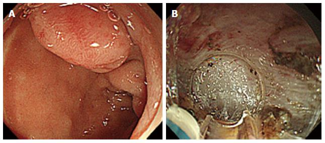
Figure 1 Endoscopic submucosal dissection of a neuroendocrine tumors in the superior duodenal bulb.
A: A protruded-type tumor 0.9 cm × 0.9 cm in size was identified; B: We performed a submucosal dissection. The tip of a knife is perpendicularly oriented to the dissection surface.
Figure 2 Endoscopic submucosal dissection of an adenoma in the descending part of the duodenum.
A: A depressed type tumor 1.2 cm × 1.2 cm in size was identified; B: After incising the oral side of the lesion, we slightly detached it to form a mucosal flap; C: A severe submucosal fibrosis was found.
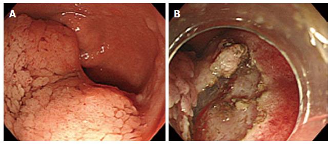
Figure 3 Endoscopic submucosal dissection of an adenoma in the anterior duodenal bulb.
A: A flat-elevated type tumor 6.0 cm × 5.0 cm in size was identified; B: We performed submucosal dissection using the ST hood.
- Citation: Matsumoto S, Miyatani H, Yoshida Y. Future directions of duodenal endoscopic submucosal dissection. World J Gastrointest Endosc 2015; 7(4): 389-395
- URL: https://www.wjgnet.com/1948-5190/full/v7/i4/389.htm
- DOI: https://dx.doi.org/10.4253/wjge.v7.i4.389


