Copyright
©The Author(s) 2015.
World J Gastrointest Endosc. Apr 16, 2015; 7(4): 354-363
Published online Apr 16, 2015. doi: 10.4253/wjge.v7.i4.354
Published online Apr 16, 2015. doi: 10.4253/wjge.v7.i4.354
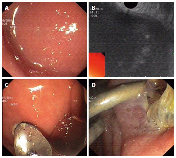
Figure 1 Endoscopic view of intragastric bulge due to pancreatic fluid collections (A), endosonographic view of pancreatic fluid collections (B), Dilation of fistula with Controlled radial expansion (CRETM) catheter balloon (C), Placement of double pigtail plastic stents through the fistula (D).
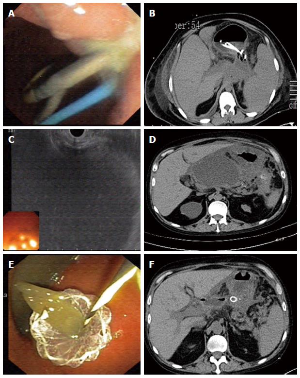
Figure 2 Placement of double pigtail plastic stent and nasocystic drain (A), computed tomography view of pancreatic fluid collections after insertion of stent and nasocystic drain (B), endosonographic view of pancreatic fluid collections before drainage (C), computed tomography view of pancreatic fluid collections before drainage (D), Placement of NAGI stent into pancreatic fluid collections (E), computed tomography view after placement of NAGI stent (F).
- Citation: Puri R, Thandassery RB, Alfadda AA, Kaabi SA. Endoscopic ultrasound guided drainage of pancreatic fluid collections: Assessment of the procedure, technical details and review of the literature. World J Gastrointest Endosc 2015; 7(4): 354-363
- URL: https://www.wjgnet.com/1948-5190/full/v7/i4/354.htm
- DOI: https://dx.doi.org/10.4253/wjge.v7.i4.354


