Copyright
©The Author(s) 2015.
World J Gastrointest Endosc. Mar 16, 2015; 7(3): 265-273
Published online Mar 16, 2015. doi: 10.4253/wjge.v7.i3.265
Published online Mar 16, 2015. doi: 10.4253/wjge.v7.i3.265
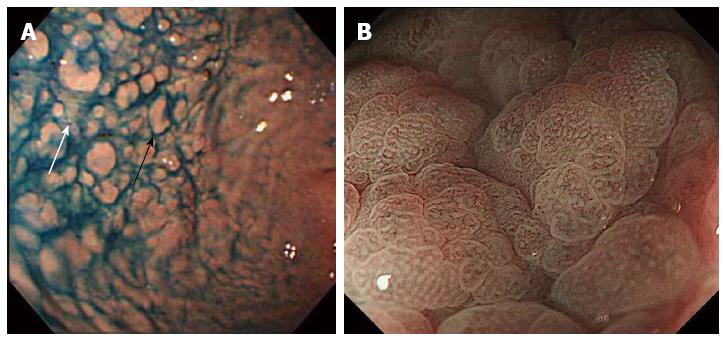
Figure 1 Endoscopic findings of collagenous gastritis.
A: Nodular lesions (black arrow) in the greater curvature of the gastric body. Depressive mucosal lesions are seen in between nodular lesions (white arrow)[34]; B: Magnifying endoscopic image with narrow band imaging. Amorphous or absent surface pit pattern and abnormal capillary vessel patterns are seen in the depressed mucosal area[41].
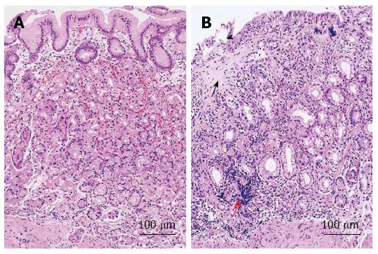
Figure 2 Histological findings of collagenous gastritis.
A: Nodular mucosal lesion did not show marked inflammatory infiltration and collagen deposition; B: Depressive mucosal lesion showed a thick collagen deposition (black arrow) and inflammatory infiltrates (red arrow). The glandular atrophy and epithelial damage is marked (black arrowhead)[34].
- Citation: Kamimura K, Kobayashi M, Sato Y, Aoyagi Y, Terai S. Collagenous gastritis: Review. World J Gastrointest Endosc 2015; 7(3): 265-273
- URL: https://www.wjgnet.com/1948-5190/full/v7/i3/265.htm
- DOI: https://dx.doi.org/10.4253/wjge.v7.i3.265


