Copyright
©The Author(s) 2015.
World J Gastrointest Endosc. Mar 16, 2015; 7(3): 253-257
Published online Mar 16, 2015. doi: 10.4253/wjge.v7.i3.253
Published online Mar 16, 2015. doi: 10.4253/wjge.v7.i3.253
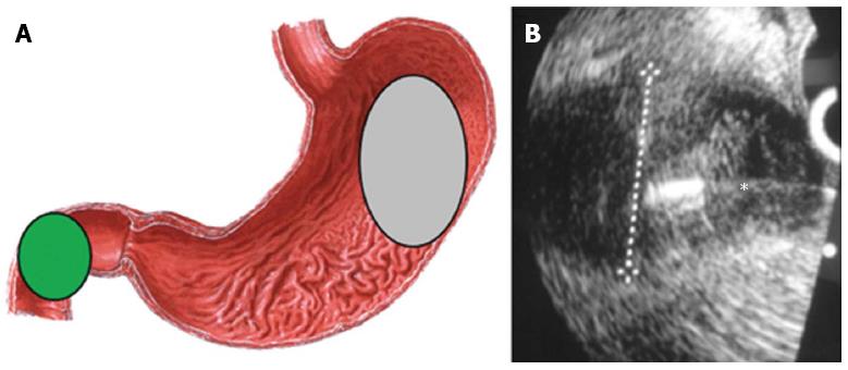
Figure 1 The right kidney image.
A: Endoscopic ultrasound positioning and access for tissue sampling; B: Fine needle aspiration of a renal lesion. The asterix is over the needle to show the fine needle aspiration of the tumor.
- Citation: Lopes RI, Moura RN, Artifon E. Endoscopic ultrasound-guided fine-needle aspiration for the diagnosis of kidney lesions: A review. World J Gastrointest Endosc 2015; 7(3): 253-257
- URL: https://www.wjgnet.com/1948-5190/full/v7/i3/253.htm
- DOI: https://dx.doi.org/10.4253/wjge.v7.i3.253


