Copyright
©The Author(s) 2015.
World J Gastrointest Endosc. Mar 16, 2015; 7(3): 224-229
Published online Mar 16, 2015. doi: 10.4253/wjge.v7.i3.224
Published online Mar 16, 2015. doi: 10.4253/wjge.v7.i3.224
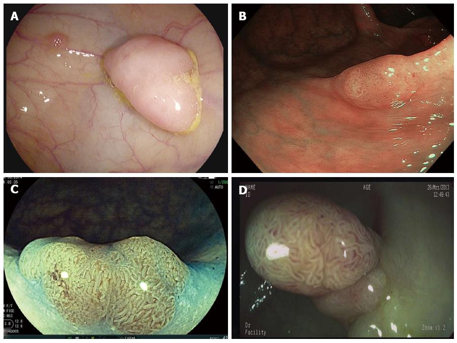
Figure 1 Colonic polyp imaged with high-definition white-light (A), narrow band imaging (B), Fujinon Intelligent Color Enhancement (C) and i-scan (D).
Data on detection rates are inconsistent. Nevertheless, dye-less chromoendoscopy techniques allow for a detailed and adequate examination of the mucosal pit pattern and the mucosal vascular pattern morphology to predict polyp histology in real time.
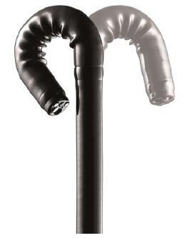
Figure 2 RetroView devices allow for a 210 degree bending of the distal tip and are equipped with virtual chromoendoscopy techniques and large working channels to allow adequate characterization of colonic lesions and subsequent endoscopic therapy (Image with kind permission from Fujifilm).
Figure 3 Full Spectrum Endoscopy-System allows 330° imaging on three contiguous monitors.
Here, a small non-polypoid lesion located on the proximal part of the ileocecal valve is only visible on the right monitor. Those lesions can easily being missed as they are often located in the realm of the shades.
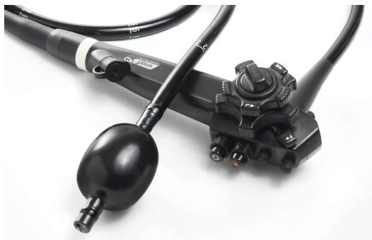
Figure 4 Newly introduced G-EYE endoscope is equipped with a balloon at the distal bending section of the endoscope.
During withdrawal the balloon is inflated thereby stabilizing the endoscope for subsequent therapeutic maneuvers. In addition the balloon yields in a straightening of the colonic folds thereby potentially improving adenoma detection rates (Image with kind permission from Smart Medical).
- Citation: Neumann H, Nägel A, Buda A. Advanced endoscopic imaging to improve adenoma detection. World J Gastrointest Endosc 2015; 7(3): 224-229
- URL: https://www.wjgnet.com/1948-5190/full/v7/i3/224.htm
- DOI: https://dx.doi.org/10.4253/wjge.v7.i3.224


