Copyright
©The Author(s) 2015.
World J Gastrointest Endosc. Mar 16, 2015; 7(3): 169-182
Published online Mar 16, 2015. doi: 10.4253/wjge.v7.i3.169
Published online Mar 16, 2015. doi: 10.4253/wjge.v7.i3.169
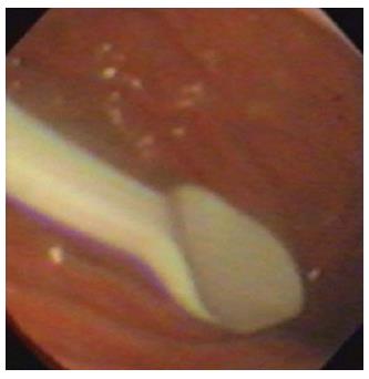
Figure 1 Foreign body (a plastic spoon) in the stomach of a child.
Ingestion of coins and small lithium batteries tend to be much more common.
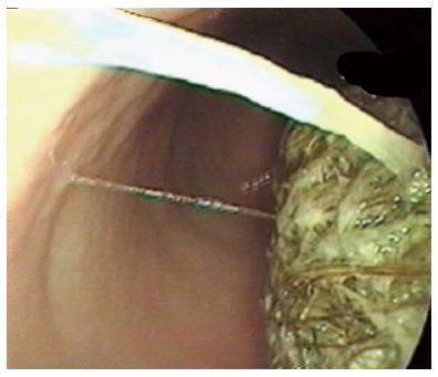
Figure 2 Bezoar seen at endoscopy.
Endoscopic removal wasn’t possible.
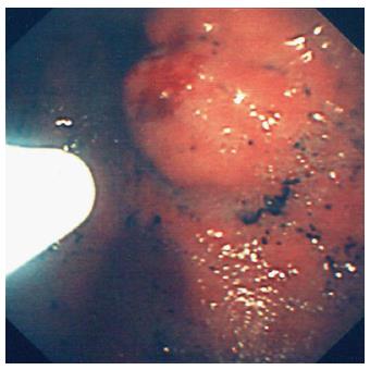
Figure 3 Injection of glue into a gastric varix.
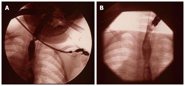
Figure 4 (A) Videofluoroscopy image of a proximal and a distal stricture in the oesophagus and (B) resolution of the strictures in the same child 3 mo after treatment with Mitomycin C.
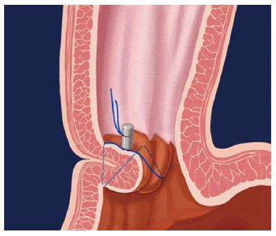
Figure 5 Endoscopic gastroplication.
This figure the pattern of a zig-zag stich when applied with an Endocinch® sewing maching.
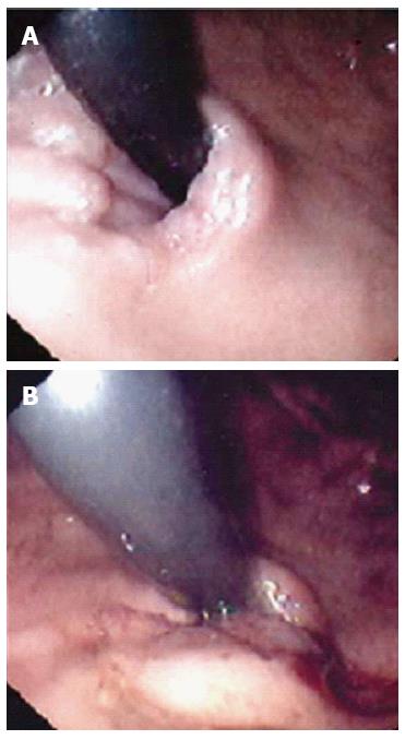
Figure 6 Endoscopic view (J manoeuvre) of a lax Gastro-Oesophageal junction in a child with major reflux before (A) and after (B) application of stitch with the EndoCinch®.
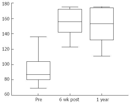
Figure 7 Significant improvement in the total QOLRAD scores (Quality of life in reflux and dyspepsia), 6 wk and 1 year after gastroplication with the Endocinch®.
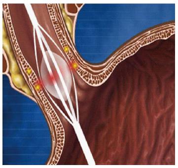
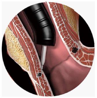
Figure 8 Use of a balloon to deliver radiofrequency energy via needle electrodes to the mucosa.
Figure 9 Injection of liquid polymer into the oesophageal mucosa.
The Enteryx® procedure.
- Citation: Rahman I, Patel P, Boger P, Rasheed S, Thomson M, Afzal NA. Therapeutic upper gastrointestinal tract endoscopy in Paediatric Gastroenterology. World J Gastrointest Endosc 2015; 7(3): 169-182
- URL: https://www.wjgnet.com/1948-5190/full/v7/i3/169.htm
- DOI: https://dx.doi.org/10.4253/wjge.v7.i3.169


