Copyright
©The Author(s) 2015.
World J Gastrointest Endosc. Dec 25, 2015; 7(19): 1318-1326
Published online Dec 25, 2015. doi: 10.4253/wjge.v7.i19.1318
Published online Dec 25, 2015. doi: 10.4253/wjge.v7.i19.1318
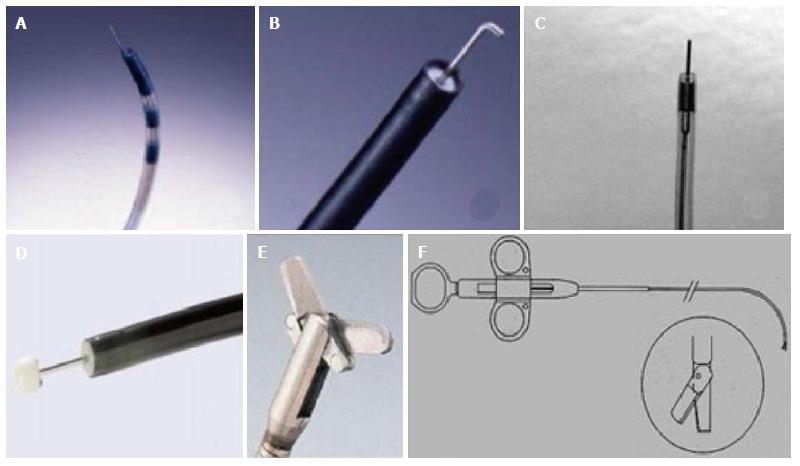
Figure 1 Accessories for incisional therapy.
A: Triple lumen needle knife; B: Hook knife; C: Needle knife (KD 10Q); D: Insulated tip knife; E: Endoscopic surgical scissors (Image courtesy of Olympus); F: Heiss-Device flexible endoscopic scissors (image courtesy of Telemed systems).
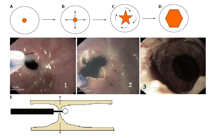
Figure 2 The technique of endoscopic incisional therapy procedure.
A-D: Schematic front view of stricture site; B: Arrows depict the radial direction of incision; C: Curved arrows depict the slicing off of the intervening areas; D: Final outcome at the end of procedure; E: Lateral view of stricture site depicting the transverse working domain of the needle knife (arrows); 1: Use of needle knife for incision; 2: After radial incision; 3: At the end of EIT and balloon dilatation. EIT: Endoscopic incisional therapy.
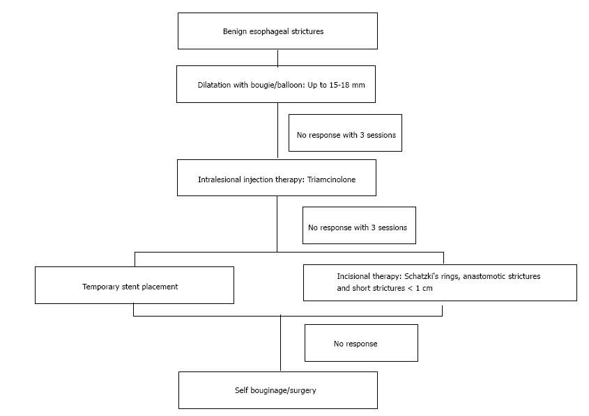
Figure 3 Algorithm for the management of benign esophageal strictures.
- Citation: Samanta J, Dhaka N, Sinha SK, Kochhar R. Endoscopic incisional therapy for benign esophageal strictures: Technique and results. World J Gastrointest Endosc 2015; 7(19): 1318-1326
- URL: https://www.wjgnet.com/1948-5190/full/v7/i19/1318.htm
- DOI: https://dx.doi.org/10.4253/wjge.v7.i19.1318


