Copyright
©The Author(s) 2015.
World J Gastrointest Endosc. Dec 10, 2015; 7(18): 1268-1278
Published online Dec 10, 2015. doi: 10.4253/wjge.v7.i18.1268
Published online Dec 10, 2015. doi: 10.4253/wjge.v7.i18.1268
Figure 1 Representative fluorescence in situ hybridization microscopic image.
Shown above are individual cells from biliary brushings showing distinct fluorescence in situ hybridization (FISH) results (arranged from lowest to highest risk of malignancy) using centromere enumeration probes (CEPs) to chromosomes 3 (red), 7 (green), 17 (aqua) and the 9p21 locus (gold). Potential FISH results include negative (two copies of each probe), trisomy 7 (≥ 10 cells with ≥ 3 CEP 7 signals and ≤ 2 signals for the other probes), tetrasomy (≥ 10 cells with four signals for all four probes), and polysomy (≥ 5 cells with ≥ 3 signals for ≥ 2 of the four probes).
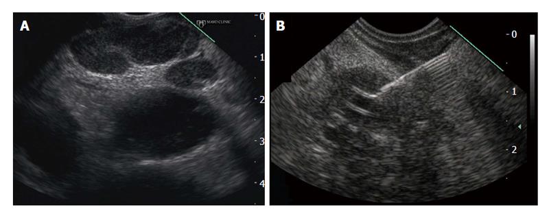
Figure 2 Endoscopic ultrasonographic findings in a patient found to have locally-advanced cholangiocarcinoma.
A: Malignant lymphadenopathy; B: Endoscopic ultrasound-guided fine needle aspiration of primary cholangiocarcinoma.
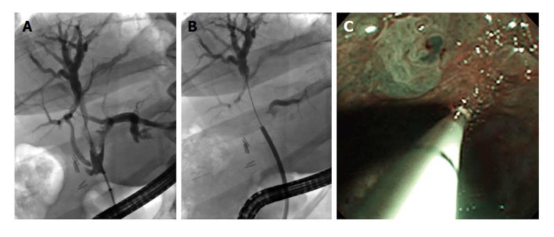
Figure 3 Passage of a SypGlass digital cholangioscope through a therapeutic duodenoscope to better evaluate hilar strictures and filling defects.
A: Hilar (primarily right anterior hepatic duct) stricture and filling defects seen during endoscopic retrograde cholangiography pancreatography; B: SypGlass cholangioscope being passed through the working channel of therapeutic duodenoscope to better assess biliary stricturing and filling defects; C: SpyGlass cholangioscopy with narrow band imaging revealing villiform biliary mucosal changes; targeted biopsies were obtained and revealed low grade dysplasia concerning for early cholangiocarcinoma.
- Citation: Tabibian JH, Visrodia KH, Levy MJ, Gostout CJ. Advanced endoscopic imaging of indeterminate biliary strictures. World J Gastrointest Endosc 2015; 7(18): 1268-1278
- URL: https://www.wjgnet.com/1948-5190/full/v7/i18/1268.htm
- DOI: https://dx.doi.org/10.4253/wjge.v7.i18.1268


