Copyright
©The Author(s) 2015.
World J Gastrointest Endosc. Nov 10, 2015; 7(16): 1208-1215
Published online Nov 10, 2015. doi: 10.4253/wjge.v7.i16.1208
Published online Nov 10, 2015. doi: 10.4253/wjge.v7.i16.1208
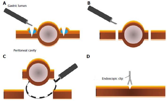
Figure 1 Illustration of the procedure for endoscopic full-thickness resection without laparoscopic assistance.
A: A circumferential incision is made to the depth of the muscularis propria around the lesion using endoscopic submucosal dissection (ESD) devices and techniques; B: After intentional perforation, the serosal layer around the lesion is incised using ESD devices; C: The tumor, including the surrounding muscularis propria and serosa, is removed using the snare; D: The gastric wall defect is closed with several metallic clips.
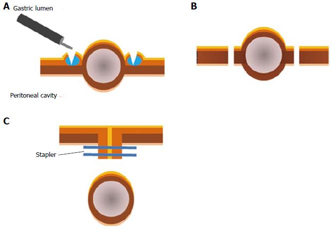
Figure 2 Illustration of the procedure for classical laparoscopic and endoscopic cooperative surgery.
A: A circumferential incision is made using endoscopic submucosal dissection (ESD) devices and techniques; B: An artificial perforation is performed from the inside of the stomach and a seromuscular incision is performed along the incision line with laparoscopic assistance. A laparoscopic incision of the remaining seromuscular layer is performed; C: The defect closure of the gastric wall is performed by laparoscopic linear staplers.
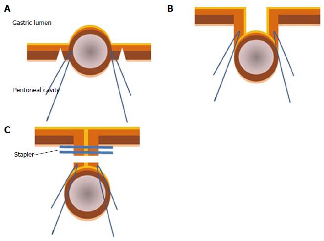
Figure 3 Illustration of the procedure for a combination of laparoscopic and endoscopic approaches to neoplasia with non-exposure technique.
A: Seromuscular layer is dissected using a laparoscopic electrocautery knife; B: Full-layer specimen is lifted by the stay sutures; C: Full-layer specimen is resected using a linear stapler.
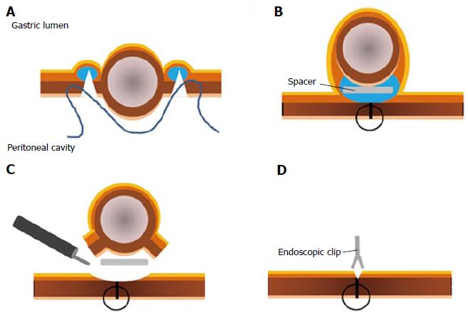
Figure 4 Illustration of the procedure for non-exposed endoscopic wall-inversion surgery.
A: circumferential seromuscular incision is performed laparoscopically outside the serosal markings after endoscopic submucosal injection; B: seromuscular layers are linearly sutured with the lesion inverted toward the inside of the stomach. A surgical sponge as a spacer is inserted between the serosal layer of the inverted lesion and the suture layer; C: Circumferential mucosal incision and the remnant submucosal incisions are made using ESD devices and techniques; D: Defect is closed with several metallic clips. ESD: Endoscopic submucosal dissection.
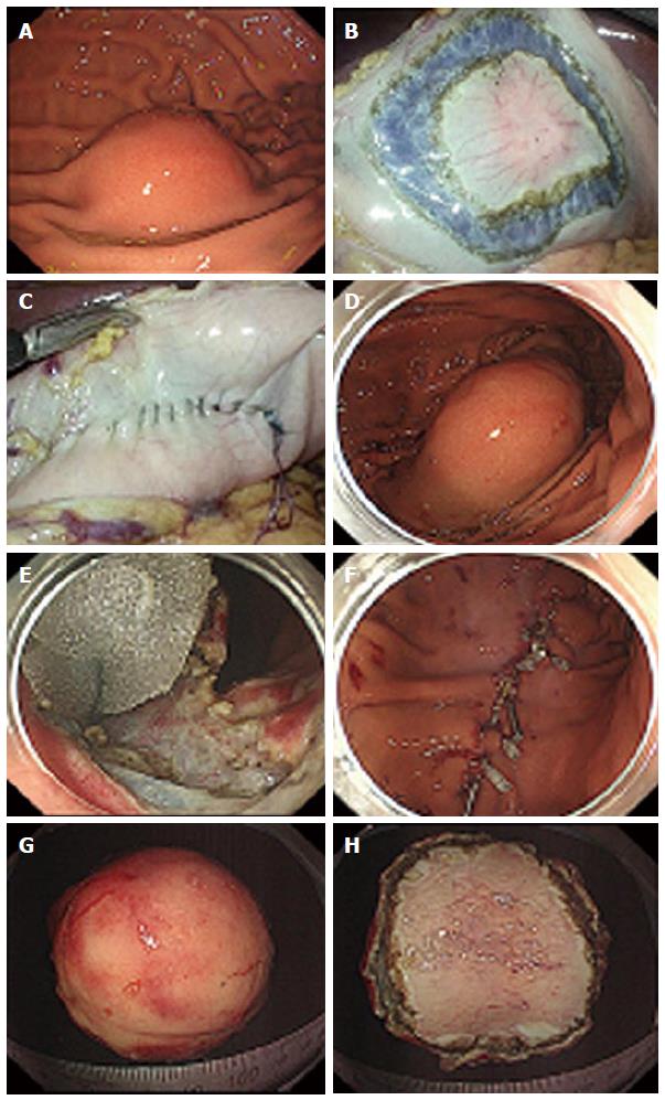
Figure 5 Procedures of non-exposed endoscopic wall-inversion surgery.
A: Protruding submucosal lesion is seen at the greater curvature of the middle gastric body; B: Circumferential seromuscular incision is made outside the serosal markings after endoscopic submucosal injection; C: Lesion is inverted with a surgical sponge used as a spacer; D: Massive protrusion of the inverted tissue; E: Surgical sponge as a spacer and a suturing line during endoscopic mucosal incision; F: Mucosal clipping after the resection; G: Resected specimen: Mucosal side; H: Resected specimen: Serosal side.
- Citation: Maehata T, Goto O, Takeuchi H, Kitagawa Y, Yahagi N. Cutting edge of endoscopic full-thickness resection for gastric tumor. World J Gastrointest Endosc 2015; 7(16): 1208-1215
- URL: https://www.wjgnet.com/1948-5190/full/v7/i16/1208.htm
- DOI: https://dx.doi.org/10.4253/wjge.v7.i16.1208


