Copyright
©The Author(s) 2015.
World J Gastrointest Endosc. Aug 25, 2015; 7(11): 1023-1031
Published online Aug 25, 2015. doi: 10.4253/wjge.v7.i11.1023
Published online Aug 25, 2015. doi: 10.4253/wjge.v7.i11.1023
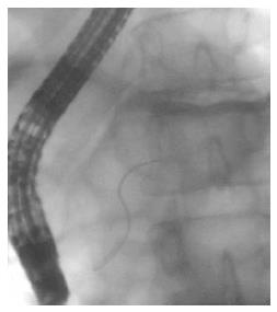
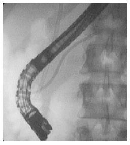
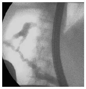
Figure 1 Endoscopic retrograde pancreatography image: Pancreas divisum with minor papilla cannulation.
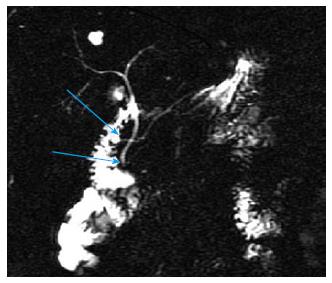
Figure 2 Secretin enhanced magnetic resonance cholangiopancreatography image: Pancreatic divisum and juxtapapillary diverticulum.
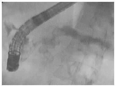
Figure 3 Endoscopic pancreatography image: Chronic pancreatitis with Wirsungolithiasis.
Figure 4 Endoscopic pancreatography image: Pancreatic duct stenosis with prestenotic dilatation after preventive pancreatic stent implantation.
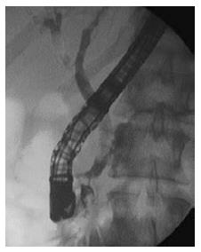
Figure 5 Endoscopic pancreatography image: Bile duct and pancreatic duct stent implantation in chronic pancreatitis.
Figure 6 Endoscopic retrograde pancreatography image: Postoperative pancreaticopleural fistula.
- Citation: Bor R, Madácsy L, Fábián A, Szepes A, Szepes Z. Endoscopic retrograde pancreatography: When should we do it? World J Gastrointest Endosc 2015; 7(11): 1023-1031
- URL: https://www.wjgnet.com/1948-5190/full/v7/i11/1023.htm
- DOI: https://dx.doi.org/10.4253/wjge.v7.i11.1023


