Copyright
©The Author(s) 2015.
World J Gastrointest Endosc. Jan 16, 2015; 7(1): 45-52
Published online Jan 16, 2015. doi: 10.4253/wjge.v7.i1.45
Published online Jan 16, 2015. doi: 10.4253/wjge.v7.i1.45
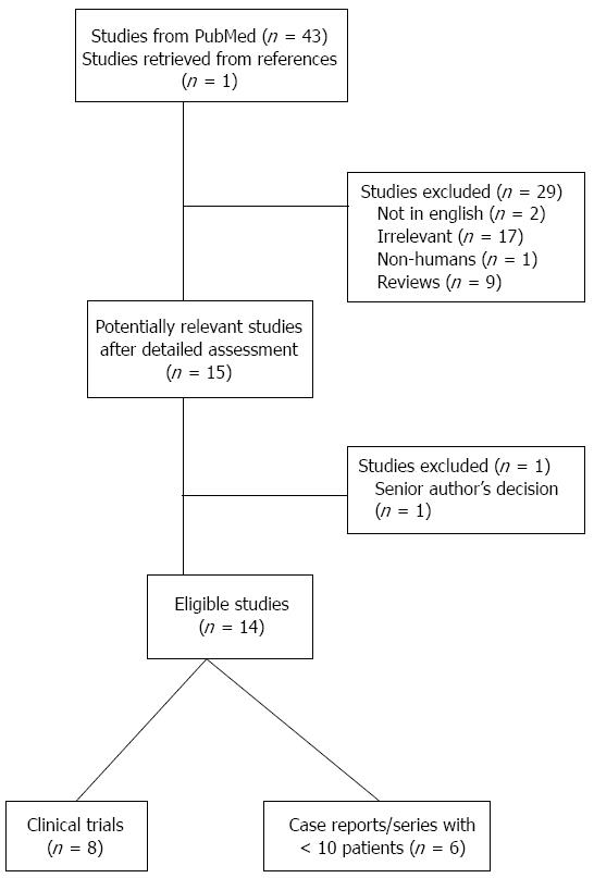
Figure 1 Flow diagram of the literature search strategy and valuation of studies identified for review.
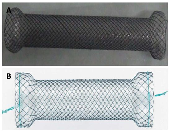
Figure 2 Large-diameter self-expandable metallic stent for achalasia.
A: Self-expandable metal stents similar to that used by Coppola et al[43]. Picture is provided by courtesy of Mr. Kuhn D, Micro-Tech Europe GmbH, Dusseldorf, Germany; B: Niti-S stent. Picture is provided by courtesy of Mr. Bekzat M, TaeWoong Medical, Seoul, South Korea.
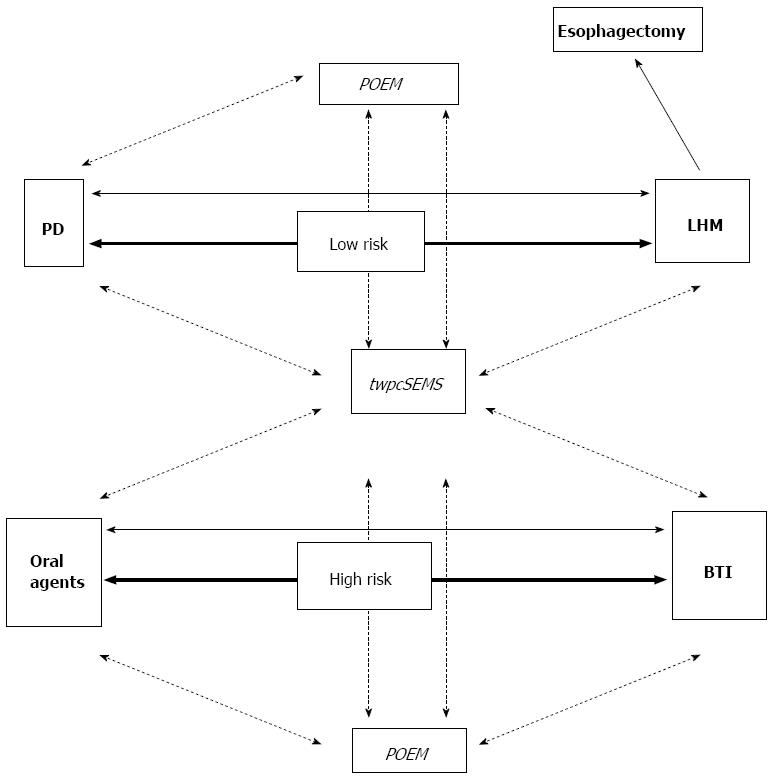
Figure 3 Proposed therapeutic algorithm for achalasia based on surgical risk.
Currently established treatments are in bold. Investigational ones are in italics. Arrows indicate current first-line treatments. Lines binding different treatments indicate management of failures. Dotted lines indicate assumed steps in management, while solid lines the to-date recommended[11]. PD: Pneumatic dilation; LHM: Laparoscopic Heller myotomy; BTI: Botulinum toxin injection; twpcSEMS: Temporal, wide, partially covered, self-expandable metallic stent; POEM: Peroral endoscopic myotomy.
- Citation: Sioulas AD, Malli C, Dimitriadis GD, Triantafyllou K. Self-expandable metal stents for achalasia: Thinking out of the box! World J Gastrointest Endosc 2015; 7(1): 45-52
- URL: https://www.wjgnet.com/1948-5190/full/v7/i1/45.htm
- DOI: https://dx.doi.org/10.4253/wjge.v7.i1.45


