Copyright
©2014 Baishideng Publishing Group Inc.
World J Gastrointest Endosc. Aug 16, 2014; 6(8): 373-378
Published online Aug 16, 2014. doi: 10.4253/wjge.v6.i8.373
Published online Aug 16, 2014. doi: 10.4253/wjge.v6.i8.373
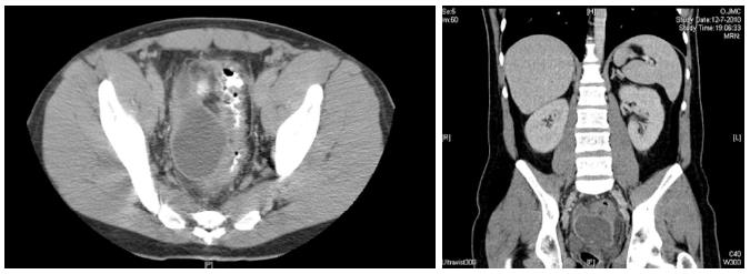
Figure 1 Axial and coronal computed tomography views of pelvic abscess adjacent to thickened sigmoid wall in a patient with acute diverticulitis.
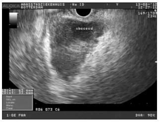
Figure 2 A pelvic abscess (43 mm × 33 mm) visualized with a linear echoendoscope (7.
5 MHz).
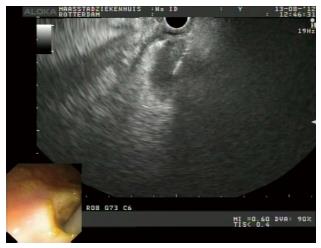
Figure 3 Endoscopic ultrasound (inlet endoscopic) image showing a cystotome used to dilate the tract.
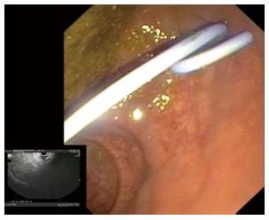
Figure 4 Endoscopic image showing the transrectal placement of 7 Fr double pig tail plastic stent.
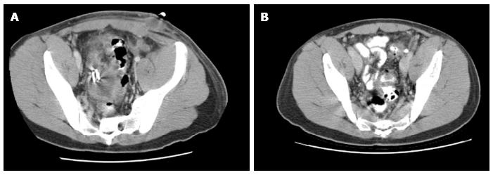
Figure 5 Follow-up computed tomography scans showing initially partial (A) and later complete (B) resolution of pelvic abscess.
- Citation: Hadithi M, Bruno MJ. Endoscopic ultrasound-guided drainage of pelvic abscess: A case series of 8 patients. World J Gastrointest Endosc 2014; 6(8): 373-378
- URL: https://www.wjgnet.com/1948-5190/full/v6/i8/373.htm
- DOI: https://dx.doi.org/10.4253/wjge.v6.i8.373


